
Analysis of BPC-157 in In-Vitro Angiogenesis
27th Oct 2026

What You Need to Know About BPC-157 Peptide
- Researchers investigate this specific peptide to understand how isolated blood vessel cells sprout, migrate, and build new capillary networks inside controlled culture dishes.
- It functions by switching on cellular surface receptors like VEGFR2 and triggering internal nitric oxide pathways that tell endothelial cells to move and link up.
- Published laboratory models consistently observe faster cell migration across artificial scratch gaps and greater capillary-like tube formation on gel matrices when this peptide is present.
- Every finding comes strictly from non-human cell cultures and preclinical models, meaning these observed cellular behaviours are for scientific investigation rather than personal use.
How BPC-157 Sparks New Vessel Formation in Cell Cultures
In laboratory dish models, BPC-157 speeds up blood vessel formation by waking up specific surface receptors and telling endothelial cells to move. It flips on internal cell switches that organise the structural scaffolding needed for tissue remodelling and capillary sprouting.
When scientists conduct an analysis of BPC-157 in in-vitro angiogenesis, they are watching how new micro-vessels sprout from existing ones inside a glass dish. Angiogenesis is the biological process that builds fresh capillary networks. Without a steady supply of nutrient-carrying tubes, recovering tissues stall out. Published studies using umbilical vein endothelial cells show that this fifteen-amino-acid sequence acts like a directional traffic controller for vessel growth.
Rather than forcing cells to divide uncontrollably, the peptide focuses on receptor availability. Specifically, it increases the expression of VEGFR2, a primary docking station on the outside of blood vessel cells. It also nudges those receptors to move from the inside of the cell out to the surface membrane where they can catch growth signals. Once those docking stations are active, the compound switches on the FAK-paxillin pathway. Think of focal adhesion kinase (FAK) and paxillin as the molecular feet of a cell. When they get phosphorylated, the cell can grip the surrounding matrix, pull itself forward, and migrate across a scratch in a culture plate.
Alongside movement, the sequence activates endothelial nitric oxide synthase, known as eNOS. This enzyme produces small amounts of nitric oxide, a gas molecule that helps newly formed tubes relax, widen, and link together into hollow loops. By coordinating receptor density, physical cell crawling, and nitric oxide output, the peptide helps scattered endothelial cells line up into organised, branching capillary structures during laboratory recovery assays.
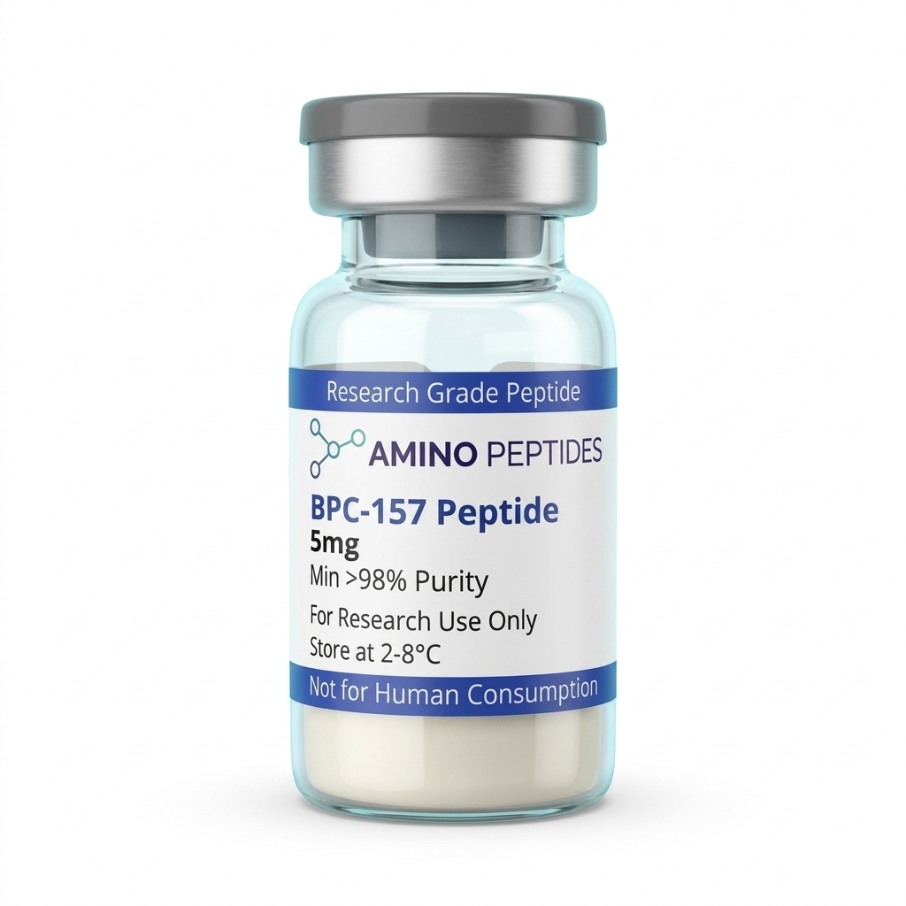
BPC-157 Peptide
From £17
Supplied as a sealed freeze-dried vial (stable 12–24 months at 2°C–8°C in a standard refrigerator).
Shop BPC-157 Peptide →Tracking the Clock from Early Signalling to Tube Networks
Watching vessel formation in a culture dish follows a clear, predictable schedule. Scientists do not wait weeks to see if a measured level of a peptide works; the earliest biochemical whispers start within minutes of contact. In the first fifteen to sixty minutes after introducing the compound to an endothelial cell monolayer, western blot tests pick up a sharp rise in phosphorylated FAK and paxillin. During this opening hour, eNOS activation also peaks, releasing nitric oxide into the liquid media. The cells look unchanged under a standard microscope at this point, yet their internal engines are already shifting gears.
Between four and twelve hours, the physical movement phase begins. Researchers often use a scratch assay, where they scrape a clean gap through a flat sheet of cells and film how fast the edges close. Cultures exposed to active protocol ratios of BPC-157 show cells detaching from their neighbours, stretching out their membranes, and crawling into the empty gap much faster than unexposed control dishes. Messenger RNA levels for VEGFR2 also climb steadily during this mid-stage window, giving the moving cells extra antennae to guide their path.
Structural remodelling becomes visible between twelve and twenty-four hours. When endothelial cells are placed on a thick gel matrix like Matrigel, they stop acting like a flat pavement and begin building three-dimensional architecture. By the eighteen-hour mark, laboratory cameras capture cells aligning end-to-end to form cord-like branches. By twenty-four to forty-eight hours, these cords hollow out and connect into polygonal mesh networks that resemble tiny capillary beds. Researchers measure the total tube length and count the branching nodes to score the final outcome. Understanding this split between rapid thirty-minute signalling and twenty-four-hour structural tube building helps investigators pick the exact moment to stop an experiment and analyse their samples.
Comparison of Angiogenic Signalling Agents in In-Vitro Endothelial Assays
| Research Compound | Molecular Classification | Primary Signalling Pathway | Observed In-Vitro Endothelial Outcome |
|---|---|---|---|
| BPC-157 | Synthetic Pentadecapeptide (15 amino acids) | VEGFR2 internalisation and VEGFR2-Akt-eNOS activation | Accelerates endothelial tube network assembly and scratch-assay migration without requiring direct VEGF binding |
| VEGF-A (Vascular Endothelial Growth Factor) | Endogenous Homodimeric Glycoprotein (~45 kDa) | Direct extracellular binding and dimerisation of VEGFR1 and VEGFR2 | Potently stimulates endothelial proliferation, sprouting, and monolayer permeability in culture |
| TB-500 (Thymosin Beta-4 Fragment) | Synthetic Polypeptide (43 amino acids / active fragment) | G-actin sequestration and focal adhesion kinase (FAK) modulation | Promotes cytoskeletal reorganisation and rapid cell motility across extracellular matrix substrates |
| bFGF (Basic Fibroblast Growth Factor) | Endogenous Signalling Polypeptide (~18 kDa) | FGFR tyrosine kinase phosphorylation and MAPK/ERK cascade | Drives robust basement membrane breakdown and three-dimensional capillary sprouting in collagen gels |
| GHK-Cu (Copper Peptide) | Tripeptide-Copper(II) Complex | Modulation of matrix metalloproteinases (MMPs) and integrins | Supports extracellular matrix remodelling and structural scaffolding around developing endothelial tubes |
Pairing BPC-157 with Complementary Sequences in the Lab
In living tissue models and advanced cell cultures, blood vessel growth rarely happens in isolation. Endothelial cells need a clear path through the extracellular matrix and plenty of structural building blocks to form stable tubes. Because of this, researchers frequently investigate how BPC-157 behaves alongside other sequences that handle different stages of the tissue remodelling process. While BPC-157 excels at turning on VEGFR2 receptors and firing up the FAK-paxillin grip mechanism, it does not directly manage the raw actin proteins that make up the internal skeleton of a cell.
That specific job belongs to Thymosin Beta-4 fragments. When investigators examine bpc157 and tb500 together in dual-agent assays, they are looking at two separate but friendly mechanisms. TB-500 binds to globular actin monomers, stopping them from clumping too early and keeping a ready pool of building blocks available at the leading edge of a moving cell. BPC-157 gives the cell the signal to move and sets up the surface anchors, while TB-500 supplies the flexible internal scaffolding to lengthen the cell body. In scratch and tube-formation models, this combination covers both the command signal and the physical hardware needed for rapid capillary bridging.
Another common partner in laboratory models is GHK-Cu, a naturally occurring copper-binding tripeptide. While the first two sequences focus on cell migration and vessel sprouting, GHK-Cu adjusts the surrounding environment by regulating metalloproteinases and collagen synthesis. New capillary tubes are fragile; they need a sturdy collagen jacket around them so they do not collapse once formed. Designing a multi-agent peptides stack allows laboratory teams to observe how receptor upregulation, actin dynamics, and extracellular matrix remodelling overlap during complex regeneration phases without overlapping on the exact same receptor pathway.
Reconstitution calculator
Choose a vial mass and a bacteriostatic reconstitution solution volume. The figure is the resulting concentration.
Concentration: 2.50 mg/mL
Per 0.01mL (10µL): 25.0 mcg
Open the full calculatorBacteriostatic reconstitution solutionWhy Analytical Purity and Cold Storage Dictate Assay Success
Cell culture assays are notoriously sensitive to chemical hitchhikers. When conducting an analysis of BPC-157 in in-vitro angiogenesis, the difference between a thriving mesh of capillary tubes and a plate of stalled, shrivelled cells often comes down to what else is hiding inside the vial. Standard solid-phase synthesis uses trifluoroacetic acid, or TFA, to cleave the finished amino acid chain from its resin bead. If a manufacturer skips proper salt-exchange steps, residual TFA stays bound to the peptide powder.
In a small glass well holding just a few millilitres of liquid media, excess TFA drops the pH and acts as a direct toxin to delicate endothelial cells. It burns through cell membranes and stalls tube formation, ruining the data before the experiment even gets going. That is why sourcing batches verified at 99%+ purity via High-Performance Liquid Chromatography (HPLC) and Mass Spectrometry is standard practice for reliable laboratory work. High-grade purification strips out truncated peptide fragments and removes harsh TFA salts, ensuring that the observed migration and VEGFR2 signalling come from the sequence itself rather than acidic contaminants.
Once a clean lyophilised powder arrives at the lab, temperature control keeps the fifteen-amino-acid chain intact. Dry vials belong in a refrigerator set between 2°C and 8°C, or in a freezer for long-term holding, away from bright light. When preparing the liquid for an assay using a sterile bacteriostatic reconstitution solution, the liquid should be trickled gently down the inside glass wall rather than sprayed straight onto the powder cake. Violent shaking shears peptide bonds and creates foam, so a slow swirl is all that is needed. Keeping reconstituted solutions cold at 2°C to 8°C prevents bacterial growth and stops the sequence from breaking down between timed assay runs.
Frequently Asked Questions
Why is an analysis of BPC-157 in in-vitro angiogenesis frequently searched?
People search for this topic because published studies show the peptide encourages new blood vessel sprouting in isolated cell cultures. Researchers analyse how it influences vascular endothelial growth factor receptor signalling during tissue remodelling. This specific cellular pathway helps scientists understand how blood flow returns to damaged areas in preclinical models.
What is the typical timeline for observing vascular changes in laboratory models?
In cell culture dishes, measurable endothelial cell migration and tube formation usually appear within twenty-four to seventy-two hours after introducing the study amount. Preclinical models examining broader tissue regeneration often track these vascular networks over seven to fourteen days to fully characterise how newly formed vessels mature and stabilise.
Why do researchers combine BPC-157 with TB-500 in cellular studies?
Scientists sometimes pair these two peptides in laboratory models to observe complementary signalling pathways during tissue recovery. While BPC-157 primarily influences vascular receptor activity and nitric oxide systems, TB-500 regulates actin, a vital cellular building protein. Studying them together helps researchers analyse whether combined protocol ratios optimise cell migration.
How do laboratories verify BPC-157 purity and manage fridge storage?
Laboratories confirm peptide purity using High-Performance Liquid Chromatography and Mass Spectrometry, ensuring the sample exceeds ninety-eight percent purity before testing. Once mixed with a bacteriostatic reconstitution solution, researchers store the liquid vial in a standard refrigerator between two and eight degrees Celsius to prevent the delicate amino acid chain from degrading.
Bibliography
- Hsieh MJ, Liu HT, Wang CN, et al. Modulatory effects of BPC 157 on vasomotor tone and the activation of Src-Caveolin-1-endothelial nitric oxide synthase pathway. Sci Rep. 2017;7:44666. View published research
- Hsieh MJ, Lee CH, Chueh HY, et al. Pro-angiogenic effect of BPC 157 is mediated via the VEGFR2-Akt-eNOS signaling pathway. J Mol Med (Berl). 2020;98(2):235-247. View published research
- Huang T, Zhang K, Sun L, et al. Body protective compound-157 enhances alkali-burn wound recovery in vivo and promotes proliferation, migration, and angiogenesis in vitro. compound Des Devel Ther. 2015;9:2485-2499. View published research
- Sikiric P, Seiwerth S, Rucman R, et al. Stable gastric pentadecapeptide BPC 157, Robert's stomach cytoprotection/adaptive cytoprotection/organoprotection, and Selye's stress response: progress and achievements. Curr Pharm Des. 2018;24(18):1990-2001. View published research
- Chang CH, Tsai WC, Lin MS, Hsu YH, Pang JHS. The promoting effect of pentadecapeptide BPC 157 on tendon outgrowth in vitro and in vivo involves growth hormone receptor expression. Molecules. 2014;19(11):19066-19077. View published research
⚠️ Research Use Only Disclaimer: All peptides and compounds are sold strictly for in-vitro laboratory research purposes only. Not intended for human or veterinary use, not a dietary supplement, and not approved to diagnose, treat, cure, or prevent any disease or condition. For use by qualified researchers in suitably equipped laboratory environments only. Amino Peptides Ltd operates in full compliance with UK MHRA regulations and applicable EU research chemical guidelines.