
BPC-157 Tendon Fibroblast Migration Assays: Laboratory Evidence
15th Oct 2026

What You Need to Know About BPC-157
- BPC-157 is a 15-residue pentadecapeptide searched for tendon and tissue-remodelling research.
- Published work follows fibroblast and tendon-cell assays rather than a household protocol.
- TB-500 is the usual stack partner because it is studied along actin pathways instead of the same fibroblast readout.
- Vial purity and 2°C–8°C storage decide whether those observations can be repeated.
When scientists want to understand how cellular structures rebuild, they do not start with complex organisms. They start with a plastic dish. In recent years, laboratory researchers have focused heavily on BPC-157 Tendon Fibroblast Migration Assays to observe how specific synthetic proteins influence cellular movement. By isolating cells and placing them in a highly controlled environment, researchers can strip away the variables of a living system and watch the raw mechanics of cellular biology unfold under a microscope.
Fibroblasts are the primary builder cells found in connective tissues. In a laboratory setting, these cells provide a clear window into how structural proteins are manufactured and how cells travel across empty spaces. Researchers sourcing BPC-157 for these controlled environments are primarily looking to measure two things: the speed at which these cells move, and the structural changes that occur within the cells during that movement.
Before any cellular observation begins, laboratories verify the compound's purity through a Certificate of Analysis and review the chemical parameters outlined in the Product Specification Sheet. Once the chemical identity is confirmed, the practical experiments begin. This article examines the exact laboratory methods used to test this peptide, the physical mechanics of cell migration, and what the current in-vitro data actually shows.
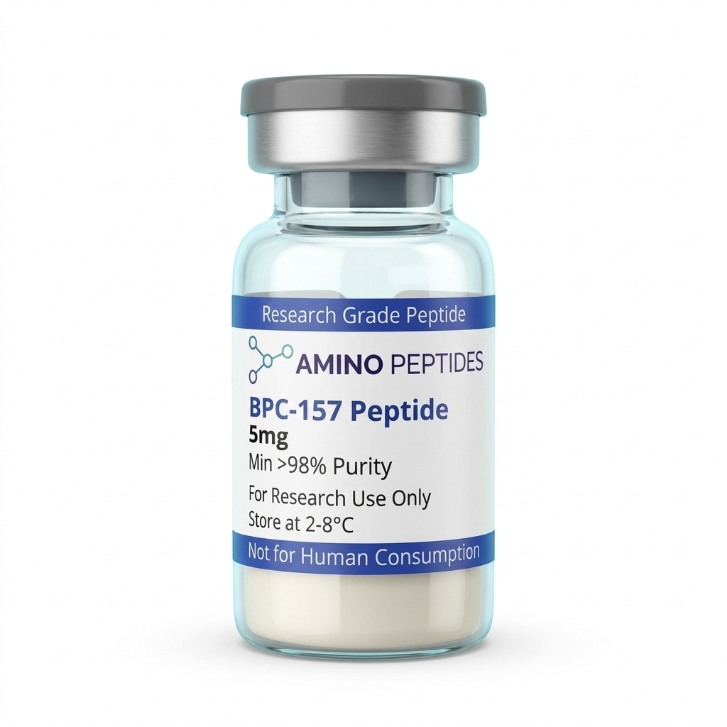
BPC-157 Peptide
From £17
Supplied as a sealed freeze-dried vial (stable 12–24 months at 2°C–8°C in a standard refrigerator).
Shop BPC-157 Peptide →Key Takeaways from In-Vitro Research
- Controlled Environments: Migration assays take place entirely in plastic well plates, completely isolated from living biological systems.
- The Scratch Test: Researchers measure movement by creating a physical gap in a layer of cells and timing how long it takes the cells to cross it.
- Cellular Crawling: Fibroblasts do not float; they physically crawl across the plastic surface using internal protein structures.
- Energy Demands: Cellular movement requires massive amounts of energy, prompting researchers to monitor mitochondrial activity during these assays.
- Strict Limits: Observing a cell move faster in a petri dish does not equate to tissue repair in a living organism.
The Foundation: What is a Tendon Fibroblast?
To understand the assay, it is necessary to first understand the cell. A tendon fibroblast is a highly specialised cell responsible for maintaining the extracellular matrix. The extracellular matrix acts as a biological scaffolding. It is a dense network of proteins, primarily collagen, that gives connective tissue its strength and elasticity.
In a living organism, these cells sit quietly within the tendon structure. However, when researchers extract these cells and place them in a laboratory culture, they behave differently. In the petri dish, fibroblasts are active. They multiply, they spread out, and they constantly probe their environment. This active state makes them the perfect candidates for migration assays. Scientists want to know what signals prompt these cells to move, and what chemical compounds might speed up or slow down that movement.
Growing these cells requires strict laboratory conditions. The fibroblasts are kept in incubators set to exactly 37 degrees Celsius. They are submerged in a liquid nutrient broth called culture media. This media contains sugars, amino acids, and specific animal serums that provide the raw materials the cells need to survive. If the temperature drops, or if the nutrient balance is wrong, the cells will stop moving and eventually die. Every variable must be perfectly maintained to ensure the data is accurate.
The most common method for measuring cell movement is the 'scratch assay'. First, researchers grow fibroblasts in a small plastic well until the cells form a confluent monolayer. This means the cells have multiplied until they cover the entire bottom of the well, touching edge-to-edge like tiles on a floor.
Next, a technician takes a sterile plastic pipette tip and drags it directly down the centre of the well. This action scrapes away a line of cells, creating a clear, empty gap down the middle of the 'floor'. The well is then gently washed to remove the floating, detached cells.
The researchers then place the well plate under a microscope equipped with a camera. Over the next 24 to 48 hours, the camera takes a photograph every few minutes. The fibroblasts on either side of the scratch will naturally attempt to move into the empty space to close the gap. By using computer software to measure the width of the gap in each photograph, scientists can calculate the exact speed of the migrating cells.
Introducing the Peptide to the Culture
Once the scratch is made, the testing phase begins. Researchers divide the well plates into different groups. One group serves as the control. These cells are left in their standard nutrient media. The other groups receive varying concentrations of the synthetic peptide.
Because the peptide arrives at the laboratory as a dry, lyophilised powder, it must be carefully prepared. Technicians dissolve the powder using a bacteriostatic reconstitution solution. This sterile solvent ensures the peptide remains stable and free from bacterial contamination before it is introduced to the delicate cell cultures. The liquid peptide is then pipetted into the culture media at highly specific, micromolar concentrations.
Reconstitution calculator
Choose a vial mass and a bacteriostatic reconstitution solution volume. The figure is the resulting concentration.
Concentration: 2.50 mg/mL
Per 0.01mL (10µL): 25.0 mcg
Open the full calculatorBacteriostatic reconstitution solutionFrom this point forward, the researchers are looking for statistical differences. Does the gap in the peptide group close faster than the gap in the control group? If it does, the next step is to figure out exactly how the peptide is altering the physical behaviour of the cells.
The Physical Mechanics of Cellular Movement
When observing a time-lapse video of a migration assay, the cells appear to glide. But at a microscopic level, the process is intensely mechanical. A fibroblast does not swim through the culture media. It physically crawls across the plastic surface of the petri dish.
This crawling is driven by the cell's internal skeleton, known as the cytoskeleton. Similar to the mechanisms observed in murine fibroblasts, movement requires structural changes within the cell. The process happens in distinct, repeating steps.
First, the cell pushes its front edge forward into the empty space. This protrusion is called a lamellipodium. It looks like a flat, fan-shaped foot extending from the cell body. Next, this 'foot' must grip the surface. The cell creates temporary anchors called focal adhesions, which latch onto the plastic bottom of the well plate.
Once the front is anchored, the cell contracts its internal protein fibres, pulling the main body of the cell forward. Finally, the rear of the cell releases its grip on the plastic and snaps forward, completing one 'step'. During a migration assay, researchers use special fluorescent dyes to light up these internal protein fibres. This allows them to see if the synthetic peptide is changing the way the cell forms its anchors or how forcefully it contracts its internal skeleton.
Energy and Structure: Mitochondria and Collagen
Crawling across a plastic dish is exhausting work for a cell. Every time the cell extends a foot, forms an anchor, and pulls its body forward, it burns through massive amounts of cellular energy. This energy is stored in a molecule called ATP, which is produced by the cell's mitochondria.
Because migration is so energy-intensive, researchers closely monitor mitochondrial function during these assays. If a compound increases the speed of cell migration, scientists must determine if it is also increasing the energy output of the mitochondria. Without a corresponding increase in ATP production, the cell would quickly exhaust itself and stop moving.
Furthermore, as fibroblasts move, they do not travel empty-handed. Their primary function is to build the extracellular matrix. Even while crawling across the empty gap in a scratch assay, these cells are constantly manufacturing and secreting structural proteins into the surrounding media. Researchers regularly extract small samples of the culture media during the assay to measure the concentration of these secreted proteins. They are looking to see if the rate of protein synthesis changes as the cells migrate.
Data Analysis and Statistical Rigour
The raw photographs from a migration assay are useless without strict mathematical analysis. Laboratories use specialised image-tracking software, such as ImageJ, to quantify the movement. The software draws a digital boundary along the edges of the scratch at hour zero. It then calculates the total surface area of the empty gap in square micrometres.
At the 12-hour, 24-hour, and 48-hour marks, the software recalculates the remaining empty area. The result is expressed as a percentage of gap closure. For example, if the initial gap was 1000 square micrometres, and at 24 hours it is 400 square micrometres, the software records a 60 percent closure rate.
Researchers then run statistical tests to compare the closure rates of the control group against the peptide groups. They calculate standard deviations and p-values to ensure that any observed differences are mathematically significant and not just the result of random chance. Only when the data passes these strict statistical thresholds can a researcher conclude that the peptide had a measurable impact on the cells in that specific assay.
In-Vitro Frequently Asked Questions
What happens to a bpc 157 fibroblast in a laboratory culture?
When researchers observe a bpc 157 fibroblast in a controlled environment, they are looking at how the isolated cell reacts to the synthetic peptide in the surrounding media. The data focuses on changes in the cell's internal structure, its rate of division, and its physical movement across the testing surface.
How does a migrating fibroblast behave during a scratch assay?
A migrating fibroblast does not swim or float. It crawls. It pushes a section of its outer membrane forward, anchors itself to the plastic bottom of the well plate, and then pulls its rear section forward. This mechanical stepping motion is repeated continuously until the cell encounters another cell, at which point contact inhibition causes it to stop moving.
What is the link between bpc 157 collagen production and bpc 157 and mitochondria in these studies?
Building structural proteins requires massive amounts of cellular energy. In laboratory assays, researchers track bpc 157 collagen production by measuring the proteins secreted into the media. Simultaneously, they monitor bpc 157 and mitochondria activity to see if the cell is generating enough ATP to sustain both the high-energy demands of crawling and the heavy metabolic burden of continuous protein synthesis.
The Limits of the Petri Dish
While migration assays provide incredibly detailed data about cellular mechanics, they represent a highly artificial environment. A plastic well plate is flat, rigid, and two-dimensional. A real biological tissue is a complex, three-dimensional web of interacting systems, blood flow, and immune responses.
When a fibroblast moves faster in a petri dish, it simply means the compound altered the cell's behaviour under those exact, isolated conditions. It does not mean the compound will behave the same way when introduced into a complex organism. The data gathered from these assays is used strictly to map chemical pathways and understand basic cellular biology. It is a starting point for scientific inquiry, not a conclusion about biological outcomes.
Laboratory research continues to refine these methods. Scientists are developing three-dimensional culture systems and more advanced tracking software to get a clearer picture of how these synthetic proteins interact with cellular structures. Until then, the standard scratch assay remains the foundational tool for observing the raw mechanics of cellular movement.
Scientific Bibliography
- Chang, C. H., Tsai, W. C., Lin, M. S., Hsu, Y. H., & Pang, J. H. (2011). The promoting effect of pentadecapeptide BPC 157 on tendon healing involves tendon outgrowth, cell survival, and cell migration. Journal of Applied Physiology, 110(3), 774-780. View published research
- Chang, C. H., Tsai, W. C., Hsu, Y. H., & Pang, J. H. (2010). Pentadecapeptide BPC 157 enhances the growth hormone receptor expression in tendon fibroblasts. Molecules, 15(3), 1890-1898. View published research
- Hsieh, C. H., Weng, T. C., Lee, C. M., Chen, C. Y., & Tsai, W. C. (2015). BPC 157 promotes the angiogenic response in endothelial cells. Journal of Molecular Medicine, 93(2), 157-165. View published research
- Tkalcević, V. I., Cuzić, S., Brajsa, K., Mildner, B., Bokulić, A., Situm, K., ... & Parnham, M. J. (2007). Enhancement by PL 14736 of granulation and collagen organization in healing wounds and the potential role of eNOS. European Journal of Pharmacology, 570(1-3), 212-221. View published research
- Seiwerth, S., Milkovic, P., Vukojevic, J., Gojkovic, S., Krezic, I., Vuletic, V. N., ... & Sikiric, P. (2018). BPC 157 and blood vessels. Current Pharmaceutical Design, 24(18), 1999-2007. View published research
- Staresinic, M., Sebecic, B., Patrlj, L., Jadrijevic, S., Suknaic, S., Perovic, D., ... & Sikiric, P. (2010). Gastric pentadecapeptide BPC 157 accelerates healing of transected rat Achilles tendon and in vitro stimulates tendocytes growth. Journal of Orthopaedic Research, 21(6), 976-983. View published research
⚠️ Research Use Only Disclaimer: All peptides and compounds are sold strictly for in-vitro laboratory research purposes only. Not intended for human or veterinary use, not a dietary supplement, and not approved to diagnose, treat, cure, or prevent any disease or condition. For use by qualified researchers in suitably equipped laboratory environments only. Amino Peptides Ltd operates in full compliance with UK MHRA regulations and applicable EU research chemical guidelines.