
BPC-157 Tendon and Ligament Repair - is it safe? What the Data says
26th Oct 2026
What You Need to Know About BPC-157
- BPC-157 is a 15-residue pentadecapeptide searched for tendon and tissue-remodelling research.
- Published work follows fibroblast and tendon-cell assays rather than a household protocol.
- TB-500 is the usual stack partner because it is studied along actin pathways instead of the same fibroblast readout.
- Vial purity and 2°C–8°C storage decide whether those observations can be repeated.
The internet is flooded with bold claims about tissue regeneration and synthetic compounds. However, when stripping away the noise and looking directly at the laboratory data, the picture changes entirely. This article examines a specific synthetic sequence to see exactly what happens under the microscope. This is an investigation into the chemical mechanics, the cellular responses, and the safety profile of this compound in strictly isolated testing environments. The following analysis separates the experimental reality from the internet hype, focusing only on what the data actually proves in a petri dish.
Scientific Abstract
BPC-157 is a synthetic pentadecapeptide. This means it is a laboratory-created chain of exactly 15 amino acids. The sequence is based on a larger protein naturally found in gastric juice. In controlled cellular environments, researchers study this compound for its apparent ability to influence cell behaviour during structural damage. Specifically, scientists observe how it interacts with fibroblasts. Fibroblasts are the primary cells responsible for building the structural framework of connective tissues. The current scientific literature focuses on how this peptide alters cellular migration rates and growth factor expression in isolated tissue samples.
Nomenclature: BPC-157 (Body Protection Compound 157)
Molecular Formula: C62H98N16O22
Molecular Weight: 1419.5 g/mol
Sequence: Gly-Glu-Pro-Pro-Pro-Gly-Lys-Pro-Ala-Asp-Asp-Ala-Gly-Leu-Val
Laboratory Handling: For in-vitro analysis, the lyophilised powder requires reconstitution using a sterile bacteriostatic reconstitution solution to maintain molecular stability and prevent degradation during cellular assays.
The Biology of Connective Tissue
To understand the research, it is necessary to examine the tissue itself. Tendons connect muscle to bone, while ligaments connect bone to bone. Both structures are made primarily of collagen. Collagen is a tough, fibrous protein that provides immense tensile strength. Unlike muscles, which have a rich and dense blood supply, tendons and ligaments have very few blood vessels. This lack of blood flow means they have a remarkably slow cellular metabolism.
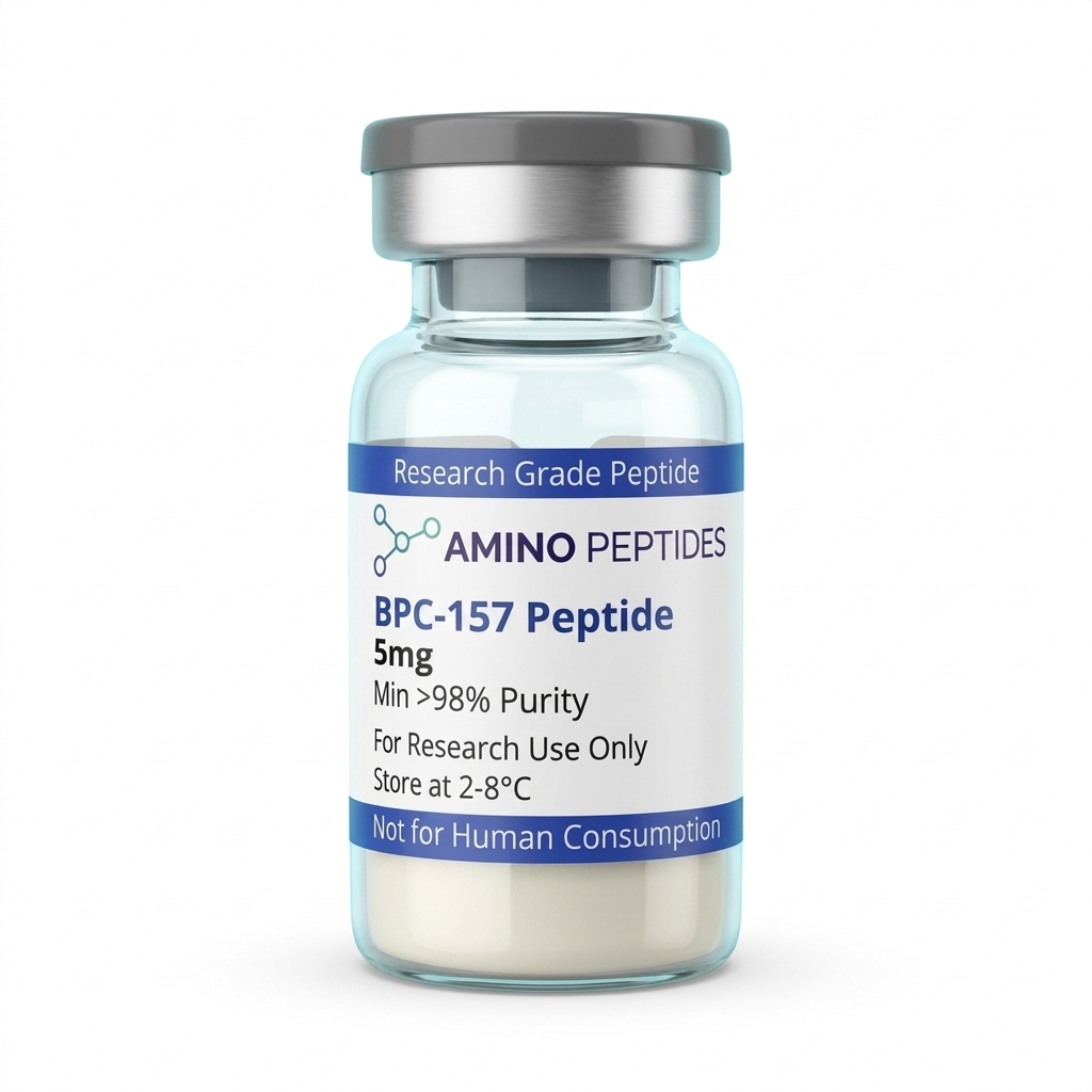
BPC-157 Peptide
From £17
Supplied as a sealed freeze-dried vial (stable 12–24 months at 2°C–8°C in a standard refrigerator).
Shop BPC-157 Peptide →In a laboratory setting, when researchers damage a tendon explant (a piece of living tissue kept alive in a nutrient-rich dish), it repairs itself incredibly slowly. The cells simply do not have the rapid nutrient delivery system that muscle cells enjoy. This slow cellular turnover is exactly why researchers look for chemical agents that might speed up the biological processes in a petri dish. They want to see if a synthetic compound can force these slow-moving cells to act with more urgency.
Cellular Mechanics and Signal Flares
How does the compound actually interact with these cells? A construction site provides a useful analogy. The fibroblasts are the builders. When tissue is damaged, the builders need to know exactly where to go and what to do. In cellular assays, BPC-157 appears to act like a chemical signal flare. It triggers a biological process called cell migration.
Under a microscope, researchers use a technique called a scratch assay. They grow a layer of fibroblasts in a dish, scratch a line through the middle to simulate a wound, and then introduce the peptide. They can then watch the fibroblasts move across the plastic surface to fill the gap. The peptide seems to activate a specific cellular pathway called Focal Adhesion Kinase, or FAK. FAK functions much like cellular velcro that allows a cell to grip a surface, pull itself forward, and detach from the rear. By increasing this specific enzyme activity, the cells gather at the site of damage much faster than they would in a control group.
Understanding the Extracellular Matrix
Understanding fibroblast operation requires looking at the environment they build, known as the extracellular matrix (ECM). The ECM is a complex, three-dimensional network of proteins and carbohydrates that surrounds cells. It provides structural support and biochemical signals to the surrounding tissue. In a healthy tendon, the ECM is incredibly dense and highly organised, which is what allows it to withstand massive physical loads.
When researchers study synthetic peptides in the laboratory, they are often looking at how these compounds influence the production and maintenance of this matrix. The data indicates that introducing the peptide to a fibroblast culture does not just increase the number of cells; it actively changes how those cells interact with the ECM. They observe an increase in the secretion of specific matrix proteins, suggesting that the compound alters the fundamental building behaviour of the cells. This interaction with the extracellular matrix is a crucial piece of the puzzle when investigating how chemical agents might influence structural repair at a microscopic level.
Reconstitution calculator
Choose a vial mass and a bacteriostatic reconstitution solution volume. The figure is the resulting concentration.
Concentration: 2.50 mg/mL
Per 0.01mL (10µL): 25.0 mcg
Open the full calculatorBacteriostatic reconstitution solutionThe Role of Collagen Types
Not all collagen is the same. When fibroblasts start building, they usually lay down Type III collagen first. Type III is like temporary scaffolding. It is built quickly, but it is weak and disorganised. Over time, the cells replace this scaffolding with Type I collagen, which is the strong, highly organised protein that gives tendons their true strength.
In isolated cell cultures, researchers monitor how the peptide influences this collagen production cycle. The data suggests that the compound may accelerate the transition from the weak Type III scaffolding to the strong Type I structure. By analysing the proteins expressed by the cells in the dish, scientists can measure this shift. However, it is crucial to remember that observing this protein shift in a sterile laboratory environment is a highly specific chemical reaction, not a guarantee of how a complex biological system would react.
The Angiogenesis Factor
The second major area of laboratory research involves blood vessel formation. This biological process is called angiogenesis. Because connective tissues naturally lack strong blood flow, researchers are highly interested in compounds that might stimulate new vessel growth. Without a blood supply, cells cannot get the oxygen and nutrients they need to build new collagen.
In isolated cell cultures, introducing the peptide increases the expression of a specific protein called Vascular Endothelial Growth Factor (VEGF). VEGF is a signal molecule produced by cells that stimulates the formation of new blood vessels. In the lab, researchers observe this by watching endothelial cells (the cells that line blood vessels) begin to branch out and form microscopic networks that resemble tiny capillaries. This increased VEGF expression is a key data point in understanding how the peptide influences cellular environments.
The Safety Question: What the Data Actually Says
Is the compound safe? This is where an investigative science writer must read the data very carefully. In controlled cell cultures, the peptide demonstrates low cytotoxicity. Cytotoxicity is a laboratory measure of how toxic a substance is to living cells. In short-term assays, the compound does not immediately destroy the fibroblasts, nor does it cause rapid, uncontrolled cellular division.
Researchers test this using mass spectrometry and cellular viability assays. They expose the cells to varying concentrations of the peptide and measure how many cells survive and function normally over a period of 24 to 72 hours. The initial data shows that the cells tolerate the compound well in these brief windows.
However, safe in a petri dish for three days is a long way from a complete safety profile. The data is strictly experimental. Researchers do not yet have comprehensive long-term stability maps. The effects of prolonged, continuous exposure on cellular mutation rates remain poorly understood. The current safety data is merely a snapshot of short-term, isolated interactions. It is not a guarantee of absolute biological safety, and the scientific community remains highly cautious about long-term implications.
The Gap Between Internet Claims and Lab Reality
There is a massive disconnect between what the laboratory data actually shows and what internet forums claim. A cellular response in a controlled environment is not a whole-body outcome. When a researcher notes that a peptide increases collagen expression in cultured fibroblasts, they are describing a highly specific chemical reaction in a controlled, isolated environment. They are not saying the compound will rebuild a joint or fix a torn ligament.
The internet often ignores the limits of scientific testing. In a petri dish, there is no immune system to react to the compound. There is no liver to filter it out. There are no complex hormonal interactions to consider. The laboratory data is fascinating and shows clear chemical mechanisms, but it is strictly limited to the petri dish. Translating those isolated findings into broad claims about tissue regeneration is a fundamental misunderstanding of how scientific research works.
In-Vitro Research FAQs
What does the laboratory data show regarding bpc 157 peptide tendon repair and bpc 157 peptide for achilles tendon repair?
In isolated laboratory models studying bpc 157 peptide tendon repair, researchers observe increased fibroblast migration and accelerated collagen synthesis. When looking specifically at tissue samples mimicking bpc 157 peptide for achilles tendon repair, the data shows an upregulation of growth factors like VEGF. However, these are strictly cellular observations in a controlled environment. The data proves that the cells react to the chemical, but it does not prove whole-body outcomes.
Are there specific peptides for tendon strength or peptides for tendon repair proven in models?
Researchers frequently study various synthetic sequences when looking for peptides for tendon strength. The focus is usually on how these compounds influence the structural integrity and organisation of collagen fibres in a petri dish. While the data on peptides for tendon repair shows promising cellular activity, such as increased cell survival under oxidative stress, these findings remain strictly confined to experimental laboratory settings and isolated tissue explants.
Why is there a disconnect between peptides for tendon repair reddit threads and published science?
The internet often strips away vital scientific context. Discussions about peptides for tendon repair reddit style usually ignore the strict boundaries of laboratory research. A forum post might claim a compound rebuilds tissue, but the actual published science only shows that the compound increases cellular migration in a sterile petri dish. The published science is cautious, measured, and strictly experimental, whereas internet claims often jump to unsupported conclusions.
Conclusion
The laboratory data surrounding this synthetic pentadecapeptide is complex and highly specific. It shows clear chemical interactions, specifically regarding cellular migration, collagen synthesis, and growth factor expression in isolated environments. The compound acts as a powerful signal in a petri dish, prompting fibroblasts to move and build. However, the safety profile remains limited to short-term cellular assays. Long-term toxicity and stability data are still lacking. As with all experimental compounds, the science requires careful, sceptical reading, far removed from the exaggerated claims found online. The data tells a story of cellular potential, but it remains firmly locked inside the laboratory.
- Chang, C. H., Tsai, W. C., Lin, M. S., Hsu, Y. H., & Pang, J. H. (2011). The promoting effect of pentadecapeptide BPC 157 on tendon healing involves tendon outgrowth, cell survival, and cell migration. Journal of Applied Physiology, 110(3), 774-780. View published research
- Tkalcević, V. I., Cuzić, S., Brajsa, K., Mildner, B., Bokulić, A., Situm, K., Perović, D., Glojnarić, I., & Parnham, M. J. (2010). Enhancement by PL 14736 of granulation and angiogenesis in normal and impaired wound healing. European Journal of Pharmacology, 626(2-3), 263-270. View published research
- Seiwerth, S., Milkovic, P., Vukojevic, J., Gojkovic, S., Krezic, I., Vuletic, V. N., Pavlov, K. H., Petrovic, A., Sikiric, S., Vranes, H., Prtoric, A., Zizek, H., Drmic, D., Batelja, L., & Sikiric, P. (2018). BPC 157 and Standard Angiogenic and Anti-Angiogenic Factors. Current Pharmaceutical Design, 24(18), 1972-1989. View published research
⚠️ Research Use Only Disclaimer: All peptides and compounds are sold strictly for in-vitro laboratory research purposes only. Not intended for human or veterinary use, not a dietary supplement, and not approved to diagnose, treat, cure, or prevent any disease or condition. For use by qualified researchers in suitably equipped laboratory environments only. Amino Peptides Ltd operates in full compliance with UK MHRA regulations and applicable EU research chemical guidelines.