
Comparing IGF-1 LR3 Kit Formats for High-Throughput Screening
15th Jul 2026
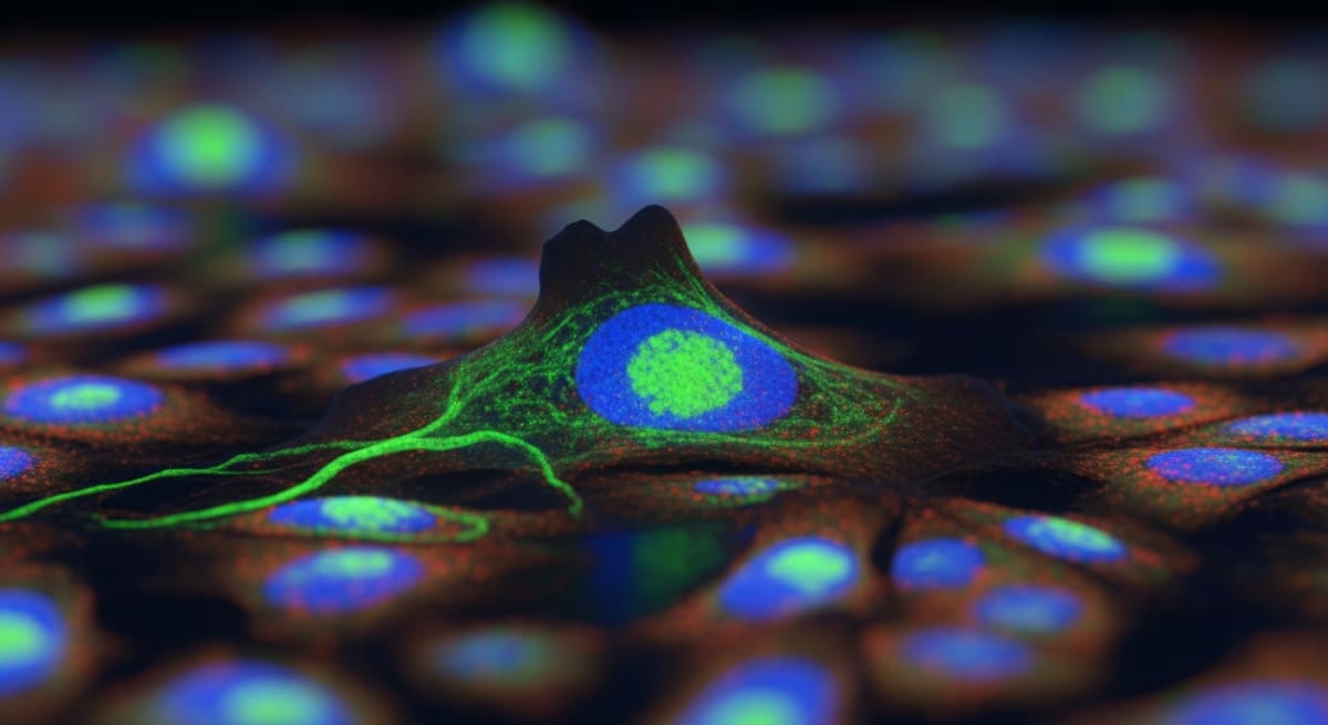
Laboratories rely heavily on Insulin-like Growth Factor 1 Long Arg3 (IGF-1 LR3) for *in-vitro* cell testing. This 83-amino-acid synthetic protein acts like natural human IGF-1, but with a structural tweak that stops binding proteins from trapping it. This modification frees the peptide to trigger cell growth and map survival pathways in isolated cell cultures. Scaling these tests into high-throughput screening (HTS) forces lab managers to make a practical choice about kit formats. The wrong setup wastes materials and ruins pipetting accuracy. When buying supplies from a reliable peptide research platform, researchers must match the product format directly to their robotic liquid handlers and plastic microplates.
Screening massive chemical libraries demands exact conditions across thousands of tiny plastic wells. A slight shift in peptide concentration or a badly mixed batch creates experimental noise. This noise easily triggers false positive results. Lab technicians must compare the physical realities of different IGF-1 LR3 formats to avoid these errors. The market offers freeze-dried powders, pre-mixed liquids, and pre-coated plates. This guide breaks down how each format handles the physical stress of automated *in-vitro* testing.
Key Takeaways
- Reconstitution Stability: Freeze-dried powders last the longest in storage. However, they demand exact robotic mixing to avoid breaking the delicate peptide chains.
- Automation Compatibility: Pre-mixed liquids cut down manual prep work. They plug straight into robotic dispensers and prevent liquid waste at the bottom of the tube.
- Assay Reproducibility: Pre-coated plates deliver the tightest consistency because they remove pipetting entirely. The trade-off is a lack of flexibility for custom dose testing.
- Binding Dynamics: The modified shape of IGF-1 LR3 stops it from sticking to natural binding proteins. This keeps the active concentration stable inside serum-free laboratory test environments.
The Molecular Profile of IGF-1 LR3
Native IGF-1 is a 70-amino-acid protein. Its use in the lab is severely limited by six specific binding proteins (IGFBPs). These proteins grab the free IGF-1 molecules and lock them away. This trapping action stops the protein from docking with the target receptor (IGF-1R) on the cell surface. As a result, scientists cannot accurately map how much protein triggers a specific cellular response.
IGF-1 LR3 beats this problem using two deliberate changes. Chemists swapped a single amino acid at position 3 and attached a 13-amino-acid tail to the front. This creates an 83-amino-acid molecule with a new physical shape. The new shape physically blocks the binding proteins from attaching, dropping their grip strength by over 120-fold. Yet, the active docking section stays completely intact. It still locks tightly onto the IGF-1R receptor on the cell membrane. Once attached, the receptor activates specific internal pathways like PI3K and MAPK/ERK. These pathways push isolated cells to multiply in serum-free liquid, matching the exact conditions used during high-throughput screening.
Tracking these growth pathways requires plotting exactly how fast different chemicals trigger a cellular reaction. For example, IGF-1 LR3 hits tyrosine kinase receptors directly to spark cell division. Laboratories mapping combined cellular reactions often test these outcomes next to ipamorelin research options. This side-by-side testing highlights how different signals change metabolic activity inside the petri dish.
High-Throughput Screening Demands and Liquid Handling
Mass compound screening uses plastic microplates stacked with 96 or 384 tiny wells. Robotic pipettes and sound-wave dispensers fire microscopic drops of testing liquid into each well. The physical traits of the peptide liquid determine whether the machines hit their targets. If the liquid is too thick or too sticky, dispensing accuracy plummets.
Peptides naturally stick to common plastics. The protein backbone clings tightly to the synthetic walls of pipette tips and storage tubes. When a machine pumps a weak peptide mixture through a plastic tip, a large chunk of the active chemical gets left behind on the plastic. This drastically lowers the actual dose hitting the test well. To fix this, lab workers add blocking agents like bovine serum albumin (BSA) to the mixing liquid. The BSA coats the plastic first, letting the active peptide slide through freely. Kit formats dictate exactly how labs handle this sticky problem.
Deep-Dive: Comparing the Three Primary Kit Formats
1. Lyophilised Monomer Vials
Freeze-dried vials offer the longest chemical shelf-life. Trapping the peptide in a vacuum inside glass vials at -80°C stops water from breaking the molecular bonds. Yet, forcing these dry powders into a robotic workflow creates practical headaches. Workers must dissolve the powder using a strict two-step method. First, a weak acid breaks down the powder. Second, a neutral buffer with a blocking protein waters it down for testing. Robots must handle this mixing step very gently. Fast, aggressive shaking whips air into the liquid. This foaming action permanently destroys the peptide chains, causing them to clump together. Pumping liquid from the mixing vial into the final plates also leaves unused liquid trapped in the tubes, wasting expensive supplies.
2. Pre-dissolved Liquid Concentrates
Pre-mixed liquid kits bypass the mixing phase entirely. The manufacturer delivers the peptide already dissolved in a clean, stable buffer fluid designed to block clumping. Technicians can drop these vials straight into the robotic handler. Skipping the mixing step deletes a major source of human and mechanical error, ensuring every test plate receives the exact same dose. The liquid mixtures carry special chemical stabilisers that keep the peptide intact at standard fridge temperatures for several weeks. This suits continuous daily testing. Their main weakness is long-term storage; sitting in water eventually cuts the peptide bonds apart, making their shelf-life much shorter than dry powders.
3. Pre-coated Multi-well Assay Plates
Pre-coated plates remove liquid peptide handling completely. The manufacturer bakes a dry, flat layer of stabilised IGF-1 LR3 directly into the bottom of the plastic wells. When the robot drops the cell mixture into the well, the dry peptide instantly absorbs the liquid and activates. This setup deletes all pipetting steps, wiping out the risk of sticky plastic tips and cross-contamination. It works brilliantly for highly repetitive standard tests. However, it strips away all experimental control. The lab is trapped using the exact dose painted into the well. The plates also demand totally dry storage, as any trapped air moisture will wreck the chemical coating.
Reconstitution and Liquid Handling Mechanics
Labs using dry powders or concentrated liquids must follow strict mixing rules. IGF-1 LR3 repels water and clumps together violently if the acid balance shifts too fast. Workers must dissolve it first with a high-acid liquid (pH 3.0 to 4.0) to break the powder down into single molecules. After it dissolves fully, they cut the harsh acid with a neutral testing liquid containing a tiny amount of BSA or Tween-20. This extra additive takes the hit from sticky plastic surfaces. It coats the machines, leaving the active IGF-1 LR3 floating freely so it can bind properly to the isolated cells during the test run.
Comparative Analysis of IGF-1 LR3 Formats
The chart below maps the practical traits of each kit format to help lab managers align their supplies with robotic screening hardware.
| Metric | Lyophilised Vials | Liquid Concentrates | Pre-coated Plates |
|---|---|---|---|
| Long-term Stability | Excellent (-20°C) | Moderate (4°C) | Good (Stored with desiccant) |
| Preparation Time | High (Requires reconstitution) | Low (Direct dilution) | Zero (Ready to use) |
| Automation Integration | Moderate (Requires robot steps) | Excellent (Deck-ready) | Excellent (No dispensing needed) |
| Experimental Flexibility | High (Custom concentrations) | High (Custom dilutions) | Low (Fixed concentration) |
| Risk of Pipetting Error | Moderate to High | Low | Negligible |
In-Vitro Scientific FAQs
Q1: Why does IGF-1 LR3 show higher potency than standard IGF-1 during cell tests involving blood serum?
Normal blood serum contains natural binding proteins that trap standard IGF-1. Once trapped, the protein cannot reach the cell receptors. IGF-1 LR3 uses a modified physical shape to block these binding proteins from attaching. Because it dodges these traps, far more of the active peptide stays free in the liquid. This free peptide successfully triggers the cellular receptors, producing a stronger reaction from a much smaller dose.
Q2: How does water break down IGF-1 LR3, and how do labs stop this during automated tests?
Water breaks the peptide apart through oxygen exposure and chemical stripping at specific amino acid points. Warm temperatures and neutral liquid levels speed up this destruction. To protect the chemical during a long robotic run, lab technicians mix the stock liquid in a slightly acidic buffer. They lock the mixing trays onto chilled robotic decks to keep the temperature low. They also block out direct light and use highly purified mixing water to stop stray enzymes from chopping the peptide apart.
Q3: Do chemical soaps in the mixing liquid skew cell-based screening results?
Labs add tiny amounts of mild chemical soaps like Tween-20 to stop peptides from sticking to plastic tubes. Cultured cells generally survive these microscopic amounts without issue. However, if a technician mixes the soap too strongly, the chemical tears holes in the delicate cell walls. This damage ruins the baseline health of the cells and creates faulty data. If a specific cell line proves too fragile for soap, labs swap it out for a harmless blocking protein like BSA.
- Francis, G. L., et al. (1992). 'Novel recombinant analogue of insulin-like growth factor-I (IGF-I), [Arg3]IGF-I, with decreased affinity for IGF-binding proteins, is more potent than IGF-I in promoting growth in vitro.' J Mol Endocrinol. View published research
- King, R., et al. (2002). 'Production and characterisation of recombinant insulin-like growth factor-I analogues with reduced affinity for binding proteins.' Biotechnol Prog. View published research
- Slaaby, R., et al. (2006). 'Ligand binding and activation of the human insulin-like growth factor-1 receptor by IGF-1 analogues.' J Biol Chem. View published research
- Yandell, C., et al. (2004). 'In vitro evaluation of IGF-1 analogues in cell culture systems for biopharmaceutical production.' Cytotechnology. View published research
⚠️ Research Use Only Disclaimer: All peptides and compounds are sold strictly for in-vitro laboratory research purposes only. Not intended for human or veterinary use, not a dietary supplement, and not approved to diagnose, treat, cure, or prevent any disease or condition. For use by qualified researchers in suitably equipped laboratory environments only. Amino Peptides Ltd operates in full compliance with UK MHRA regulations and applicable EU research chemical guidelines.