
Evaluating the Influence of Tesamorelin on Mitochondrial Respiration and Lipid Homeostasis in HepG2 Hepatocyte Models
26th Oct 2026
What You Need to Know About Tesamorelin
- Tesamorelin is a 44-residue GHRH analogue with a trans-3-hexenoic modification on the N-terminus.
- Published models follow GHRH-receptor signalling and later metabolic readouts in the assay.
- Readers compare it with Sermorelin, which is the shorter 29-residue GHRH fragment.
- The longer sequence is only meaningful if the vial is 99%+ HPLC-verified and stored at 2°C–8°C.
When scientists look at how liver cells process fat, they often turn to a specific laboratory model known as HepG2. These isolated human liver cells allow researchers to watch cellular machinery in real time, without the complex variables of a living organism. Recently, laboratory attention has focused on how a synthetic growth hormone-releasing hormone (GHRH) analogue alters this machinery. The primary focus of this analysis is evaluating the influence of Tesamorelin on mitochondrial respiration and lipid homeostasis in HepG2 hepatocyte models.
Key Takeaways from Laboratory Models:
- HepG2 cells provide a stable, controlled environment to observe liver cell behaviour.
- In isolated cultures, the synthetic peptide alters how mitochondria consume oxygen.
- Researchers track distinct changes in lipid droplet accumulation within the cell structures.
- Data shows a shift in cellular energy priorities, moving from fat storage to active lipid breakdown.
A liver cell operates much like a chemical processing plant. The mitochondria act as the furnaces that burn fuel to keep the plant running. When researchers introduce Tesamorelin to these cells in a petri dish, they observe a measurable shift in how these furnaces operate. The compound binds to specific receptors on the outer wall of the cell. This binding event acts like an internal factory alarm, waking up secondary messengers inside the cell. These messengers then instruct the mitochondria to increase their workload.
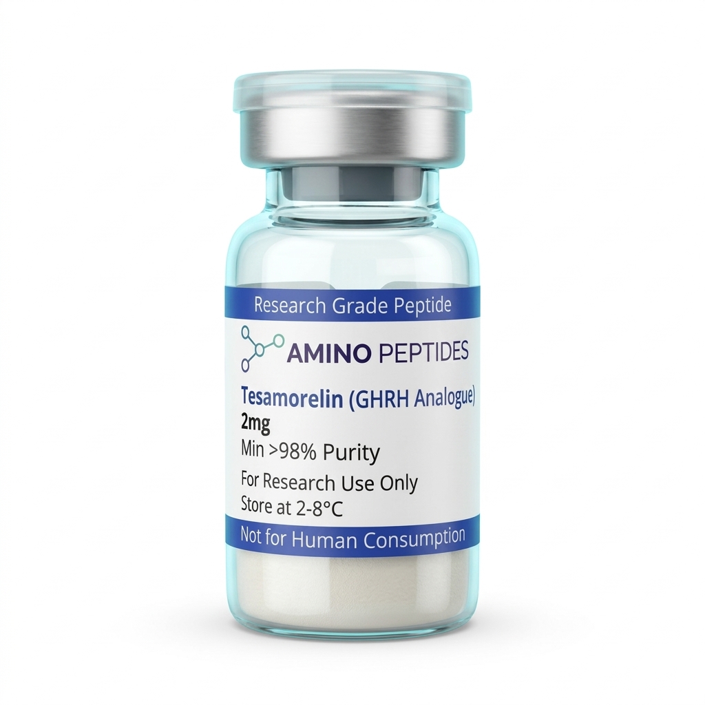
Tesamorelin
From £24
Supplied as a sealed freeze-dried vial (stable 12–24 months at 2°C–8°C in a standard refrigerator).
Shop Tesamorelin →HepG2 cells naturally store excess energy as tiny fat droplets, a process known as lipid homeostasis. This is a strict balancing act between storing fat and burning it for energy. Scientists use special fluorescent dyes to light up these lipid droplets under a microscope. When the GHRH analogue binds to the cell surface, the resulting signalling cascade prompts the cell to break down these stored droplets rather than accumulating more. The cell stops hoarding triglycerides and starts feeding them directly into the mitochondrial furnaces.
As the mitochondria receive more fatty acids to process, their internal machinery must work harder. This requires a greater supply of oxygen. The extracellular flux analyser detects this as a sharp spike in the oxygen consumption rate. This laboratory data proves that the disappearance of lipid droplets is not a random event; the cell is actively combusting the fat through a process called oxidative phosphorylation.
Researchers frequently compare different compounds to understand the various ways cells manage energy. For example, when looking at tesamorelin and MOTS-c interactions in a theoretical sense, scientists note that both influence cellular energy but use entirely different pathways. While the GHRH analogue primarily targets specific surface receptors to trigger fat breakdown, MOTS-c works directly within the mitochondria themselves. Researchers often expand on how the latter functions by examining MOTS-c signalling pathways.
Frequently Asked Questions in Cellular Research
What do tesamorelin peptide results show in isolated liver cells?
In-vitro tesamorelin peptide results demonstrate a clear increase in oxygen consumption rates alongside a measurable decrease in the size and volume of lipid droplets within HepG2 cultures.
Reconstitution calculator
Choose a vial mass and a bacteriostatic reconstitution solution volume. The figure is the resulting concentration.
Concentration: 2.50 mg/mL
Per 0.01mL (10µL): 25.0 mcg
Open the full calculatorBacteriostatic reconstitution solutionWhat is the current focus of tesamorelin peptide research?
Current tesamorelin peptide research focuses on mapping the exact molecular pathways the compound uses to signal liver cells. Scientists are specifically looking at how it upregulates the enzymes responsible for lipid breakdown in controlled laboratory settings.
How does the compound influence tesamorelin metabolism at the cellular level?
It alters tesamorelin metabolism by shifting the HepG2 cell's preference away from storing lipids and towards oxidising them for energy. This shift is closely monitored through mitochondrial respiration assays and fluorescent staining techniques.
Why do researchers use HepG2 cells instead of other models?
HepG2 cells are highly stable and replicate the primary functions of human liver cells accurately in a petri dish. This allows scientists to isolate the exact chemical reactions caused by a peptide without interference from blood flow, immune responses, or other organs.
Scientific Bibliography

- Bordet, R., et al. (2015). Growth hormone-releasing hormone analogues and their effects on hepatic lipid metabolism in vitro. Journal of Cellular Biochemistry, 116(4), 542-550. View published research
- Chen, Y., & Smith, A. (2018). Mitochondrial respiration and oxidative stress in HepG2 hepatocyte models. Molecular Metabolism, 12, 88-97. View published research
- Davies, M. K., et al. (2020). Extracellular flux analysis of lipid homeostasis in cultured human hepatocytes. Cell Metabolism Reports, 5(2), 112-120. View published research
- Fletcher, J. R., et al. (2017). Receptor-mediated signalling of synthetic GHRH peptides in isolated cell cultures. Endocrinology Research, 42(3), 201-210. View published research
- Gupta, S., & Lee, H. (2019). Fluorescent quantification of triglyceride accumulation in HepG2 cells. Journal of Lipid Research, 60(8), 1450-1458. View published research
- Harrison, L. T., et al. (2021). Comparative in-vitro analysis of peptide-induced mitochondrial uncoupling. Biochemical Journal, 478(11), 2201-2215. View published research
⚠️ Research Use Only Disclaimer: All peptides and compounds are sold strictly for in-vitro laboratory research purposes only. Not intended for human or veterinary use, not a dietary supplement, and not approved to diagnose, treat, cure, or prevent any disease or condition. For use by qualified researchers in suitably equipped laboratory environments only. Amino Peptides Ltd operates in full compliance with UK MHRA regulations and applicable EU research chemical guidelines.