
Investigating KPV's Molecular Interactions with Cellular Proliferation Pathways In-Vitro
24th Jul 2026

What You Need to Know About KPV Peptide
- Researchers look into KPV because this tiny three-amino-acid fragment shows strong cellular calming effects in laboratory dishes without triggering skin pigment changes.
- In plain English, KPV hitchhikes into cells using a specialised transporter called PepT1 and helps quieten down overactive stress signalling pathways.
- Published laboratory studies observed that adding KPV at nanomolar concentrations supports normal cell division and tissue remodelling across gut and skin cell lines.
- Unlike the full-length parent hormone alpha-MSH, this short sequence does not strongly activate surface pigment receptors, making it a focused tool for studying cellular recovery.
How the KPV Tripeptide Calms Cellular Stress to Support Tissue Recovery
KPV supports cellular recovery in laboratory models by entering cells through PepT1 transporters and quietening the NF-kappaB inflammatory switch. By lowering this internal stress noise, the tiny three-amino-acid sequence allows epithelial and dermal cells to return to normal division and barrier repair.
At the structural level, KPV is made of just three linked amino acids: lysine, proline, and valine. It is the active C-terminal tail clipped from a larger natural hormone called alpha-melanocyte-stimulating hormone, or alpha-MSH. While the parent molecule triggers pigment changes across skin cells, this tiny three-part fragment leaves the pigment switches alone and focuses purely on calming overactive cellular stress responses.
Published studies investigating KPV's molecular interactions with cellular proliferation pathways in-vitro show that its tiny size is its biggest advantage. Instead of waiting solely for surface receptors like MC1R or MC3R, KPV hitches a ride directly inside the cell using the PepT1 di- and tripeptide transporter. Once inside the cytoplasm, it blocks the import of inflammatory messengers into the cell nucleus. Specifically, it stops a protein complex called NF-kappaB from switching on genes that produce tissue-damaging cytokines such as TNF-alpha and interleukin-6.
Why does calming inflammation matter for cellular proliferation and skin elasticity? When cells in a culture dish are flooded with inflammatory signals, they lock into a defensive pause. They stop dividing, slow down collagen upkeep, and let tight junctions between neighbouring cells loosen. By quieting that inflammatory alarm, KPV clears the roadblock so normal proliferation pathways—like controlled MAPK and ERK signalling—can resume their routine maintenance work.
Importantly, in-vitro assays show that KPV does not force reckless or runaway cell division. Instead, it restores the baseline proliferation rate that healthy keratinocytes and gut lining cells use to close microscopic gaps and maintain a tight, resilient tissue barrier.
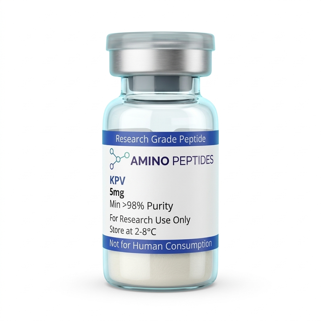
KPV Peptide
From £25
Supplied as a sealed freeze-dried vial (stable 12–24 months at 2°C–8°C in a standard refrigerator).
Shop KPV Peptide →What Cell Culture Clocks Show From Early Signalling to Barrier Renewal
When scientists observe KPV in cell culture dishes, the biological response unfolds in two clear stages: rapid intracellular signalling followed by slower structural tissue remodelling. Understanding this two-speed clock helps explain how a simple tripeptide guides recovery in preclinical models without overstimulating the cells.
During the first 30 minutes to four hours after introducing a measured level of KPV to cultured intestinal or skin cells, the action is purely chemical. The PepT1 transporter rapidly pulls the tripeptide across the cell membrane. Inside the cell, KPV stabilises the inhibitor protein I-kappaB-alpha, which keeps NF-kappaB trapped in the cytoplasm. Within two to six hours, laboratory assays measuring messenger RNA show a sharp drop in pro-inflammatory signals like IL-8 and TNF-alpha. At this early mark, the cells look exactly the same under a microscope, even though their internal stress chemistry has already quietened down.
The second stage—visible proliferation and structural recovery—takes place between 12 and 72 hours. In classic scratch-wound assays, where researchers create a clean gap across a flat sheet of cultured keratinocytes or colonic epithelial cells, KPV-exposed dishes begin showing coordinated cell migration and division around the 18-to-24-hour mark. Because the inflammatory brake has been lifted, cells along the edge of the gap multiply at a steady, healthy pace to fill the open space.
By 48 to 72 hours, published studies report measurable rebounds in transepithelial electrical resistance, a standard lab test that checks how tightly cells are zipped together. Proteins like zonula occludens-1 (ZO-1) and occludin reorganise along the cell borders, sealing the barrier. In dermal fibroblast cultures observed over three to five days, this calm environment also supports steady extracellular matrix upkeep, preserving the structural proteins that give skin models their bounce and elasticity.
Comparison of KPV and Related Research Peptides in Laboratory Models
| Property or Pathway | KPV (Lys-Pro-Val) | Full-Length alpha-MSH | KDPT (Related Tripeptide) |
|---|---|---|---|
| Amino Acid Length | 3 amino acids (tripeptide) | 13 amino acids (tridecapeptide) | 3 amino acids (synthetic derivative) |
| Primary Entry or Receptor Route | Intracellular uptake via PepT1 transporter | Binds surface melanocortin receptors (MC1R to MC5R) | Interacts with IL-1beta signalling without MC1R binding |
| Pigment Signalling (MC1R Activity) | Negligible in standard cell assays | High activation of pigment pathways | None observed in published cell models |
| Effect on NF-kappaB Signalling | Stabilises IkappaB-alpha to quieten nuclear translocation | Reduces pathway activity via cAMP second messengers | Dampens IL-1beta-driven NF-kappaB activation |
| Observed In-Vitro Proliferation Role | Restores normal epithelial cell division under stress | Modulates keratinocyte and melanocyte growth | Supports barrier recovery in stressed epithelial layers |
Pairing KPV With Complementary Peptides in Multi-Pathway Lab Models
Because KPV works mainly by quieting internal stress signals rather than pushing growth receptors directly, researchers frequently combine it with structural or antimicrobial peptides in multi-agent laboratory protocols. Pairing peptides that use separate cellular doorways lets investigators study how tissue recovery works when inflammation control and structural rebuilding happen at the same time.
One of the most studied combinations in barrier research pairs KPV with BPC-157. While KPV enters via PepT1 to calm NF-kappaB inside the cell, BPC-157 works on focal adhesion kinase (FAK) and paxillin pathways to guide cell migration and blood vessel sprouting. In combined cell culture models, KPV clears the inflammatory static while BPC-157 organises the physical scaffolding needed for fibroblasts and epithelial sheets to bridge larger gaps.
Another widely explored pairing in dermal and extracellular matrix studies combines KPV with the copper tripeptide GHK-Cu. GHK-Cu modulates hundreds of genes tied to collagen synthesis, elastin production, and antioxidant defence. When studied alongside a verified kpv peptide uk reference standard, researchers can observe how keeping cytokine levels low prevents matrix metalloproteinases (MMPs) from chewing up the fresh collagen and elastin fibres that GHK-Cu helps cells produce.
In innate immune and surface barrier research, scientists also examine KPV and LL-37 in parallel or combined assays. LL-37 is a cathelicidin peptide that disrupts unwanted microbial membranes and recruits repair signals, but high study amounts of LL-37 alone can sometimes stir up local inflammatory signalling in sensitive cell lines. Adding KPV to the protocol ratio helps temper that secondary cytokine spike, allowing researchers to study antimicrobial surface defence alongside calm, orderly epithelial cell proliferation.

Figure: Investigating KPV's Molecular Interactions with Cellular Proliferation Pathways In-Vitro
Reconstitution calculator
Choose a vial mass and a bacteriostatic reconstitution solution volume. The figure is the resulting concentration.
Concentration: 2.50 mg/mL
Per 0.01mL (10µL): 25.0 mcg
Open the full calculatorBacteriostatic reconstitution solutionWhy 99%+ HPLC Purity and TFA Removal Matter for Clean Cell Assays
When investigating KPV's molecular interactions with cellular proliferation pathways in-vitro, the quality of the lyophilised powder in the vial directly controls the accuracy of the readout. Cultured cells sit in a closed dish with no liver or kidneys to filter out manufacturing leftovers, meaning even tiny chemical impurities can skew proliferation numbers or mimic cellular stress.
The biggest hidden variable in poorly finished peptides is trifluoroacetic acid, or TFA. During solid-phase synthesis, manufacturers use TFA to cleave the finished peptide chain from its resin bead. If the lab skips secondary ion-exchange salt removal, residual TFA stays bound to the lysine amino acid in KPV. In a cell culture dish, free TFA shifts the pH of the growth medium and acts as a mild toxin to delicate keratinocytes and fibroblasts. It can slow down cell division by 20 to 30 percent, tricking an observer into thinking the peptide itself stalled proliferation when the culprit was actually leftover acid in an unrefined vial.
This is why serious preclinical work relies on batches verified at 99% or higher purity via High-Performance Liquid Chromatography (HPLC) alongside Mass Spectrometry (MS) identity confirmation, with TFA swapped out for benign acetate or hydrochloride salts. Truncated sequences—unfinished two-amino-acid scraps left behind by rushed purification—also compete for the PepT1 transporter without delivering any NF-kappaB calming activity, muddying concentration-response curves.
Proper storage keeps a high-purity batch stable from the first assay to the last. Lyophilised KPV vials should be kept in a dark refrigerator between 2°C and 8°C, or frozen at -20°C for long-term archiving. Once mixed with a sterile bacteriostatic reconstitution solution, the liquid vial must stay refrigerated at 2°C to 8°C and away from UV light to prevent oxidation or peptide bond breakdown over subsequent weeks of laboratory testing.
Frequently Asked Questions
Why are researchers investigating KPV's molecular interactions with cellular proliferation pathways in-vitro?
Researchers study KPV in cell cultures to understand how this small peptide influences cellular signalling and tissue remodelling without causing unwanted pigment changes. By observing isolated cells, scientists can clearly analyse how KPV calms inflammatory markers and supports structural recovery in controlled laboratory models.
What is a typical timeline for observing KPV in cellular proliferation assays?
In published studies, initial changes in cellular signalling often appear within six to twenty-four hours of applying a measured level of KPV. Full observations of cell division and tissue repair markers typically take between forty-eight and seventy-two hours inside controlled laboratory incubators.
Can KPV be combined with other peptides during in-vitro research?
Yes, laboratory protocols frequently pair KPV with other research peptides like BPC-157 or TB-500 to observe combined effects on tissue regeneration. Researchers carefully adjust the protocol ratio of each compound to study how different signalling pathways interact during cellular recovery experiments.
What purity levels and storage conditions are required for KPV in the laboratory?
For reliable in-vitro data, KPV should have a verified purity of at least ninety-eight percent, confirmed via high-performance liquid chromatography. Once mixed with a bacteriostatic reconstitution solution, the liquid peptide must be stored in a laboratory refrigerator at two to eight degrees Celsius.
Bibliography
- Dalmasso G, et al. PepT1-mediated tripeptide KPV uptake reduces intestinal inflammation. Gastroenterology. 2008;134(1):166-178. View published research
- Brzoska T, et al. Alpha-melanocyte-stimulating hormone and related tripeptides: biochemistry, antiinflammatory and protective effects in vitro and in vivo. Endocr Rev. 2008;29(5):581-602. View published research
- Kannengiesser K, et al. Melanocortin-derived tripeptide KPV has anti-inflammatory potential in murine models of inflammatory bowel disease. Inflamm Bowel Dis. 2008;14(3):324-331. View published research
- Luger TA, Brzoska T. Alpha-MSH related peptides: a new class of anti-inflammatory and immunomodulatingelligent agents. Ann Rheum Dis. 2007;66(Suppl 3):iii52-iii55. View published research
See our UK peptide research guides & COA briefings first in Google Search.
Add Amino Peptides UK as a Preferred Source on Google⚠️ Research Use Only Disclaimer: All peptides and compounds are sold strictly for in-vitro laboratory research purposes only. Not intended for human or veterinary use, not a dietary supplement, and not approved to diagnose, treat, cure, or prevent any disease or condition. For use by qualified researchers in suitably equipped laboratory environments only. Amino Peptides Ltd operates in full compliance with UK MHRA regulations and applicable EU research chemical guidelines.