
MOTS-C Amount: See What The Laboratory Data Actually Shows
7th Sep 2026

What You Need to Know About MOTS-C
- Curious readers often search for MOTS-c study amounts to understand how researchers measure this peptide when investigating cellular energy and stamina.
- Unlike most proteins encoded in the cell nucleus, MOTS-c comes directly from the mitochondria and acts as a natural messenger that helps optimise how cells process fuel.
- It works in plain terms by activating the AMPK energy sensor, which encourages skeletal muscle cells to pull in glucose and burn fatty acids more efficiently.
- Published studies in preclinical models have observed improved metabolic balance, stronger physical endurance on treadmills, and greater resilience against age-related cellular stress.
How MOTS-c Drives Cellular Energy in Preclinical Models
Published studies show that MOTS-c is a sixteen-amino-acid peptide encoded directly inside mitochondrial DNA rather than the cell nucleus. In preclinical models, it acts as a metabolic messenger that helps cells manage glucose uptake and energy production under stress.
Most peptides studied in modern laboratories are built from instructions stored in the main cell nucleus. MOTS-c is different. Its genetic code lives inside the mitochondria, the tiny power plants that generate cellular energy. When a cell faces metabolic stress, such as high glucose levels or intense energy demand, MOTS-c travels from the mitochondria into the nucleus to help adjust how the cell uses fuel.
In laboratory models, researchers observe that MOTS-c targets the folate-methionine cycle. By slowing down a specific step in this pathway, it causes a natural compound called AICAR to build up inside the cell. This buildup switches on AMPK, an enzyme that acts like a fuel gauge for cellular energy. Once AMPK is active, the cell starts pulling in more glucose and burning fatty acids more efficiently instead of storing them.
When people search online for a standard protocol ratio or measured level, they are usually trying to understand how much of this peptide triggers AMPK activation in published studies. In cell cultures and rodent models, scientists do not use a single universal concentration. Instead, the study amount is scaled by body mass in animal models or by micromolar concentration in isolated tissue dishes. Examining how these specific concentrations work in a dish helps explain why mitochondrial signalling is such a popular topic in longevity and metabolic research.
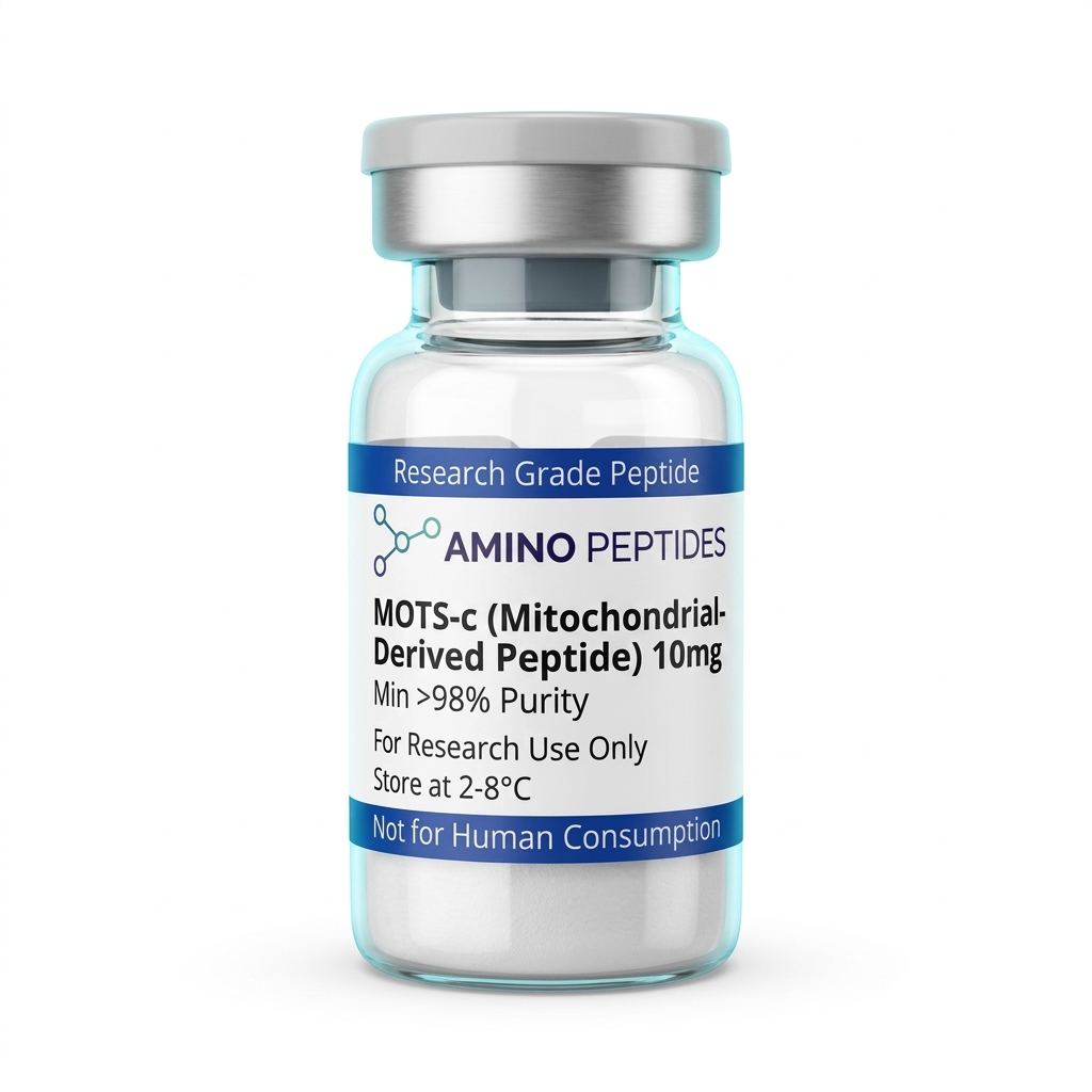
MOTS-C
From £30
Supplied as a sealed freeze-dried vial (stable 12–24 months at 2°C–8°C in a standard refrigerator).
Shop MOTS-C →What Published Studies Track Across Different Study Amounts
In published studies, the observed effects of MOTS-c depend heavily on both the measured level applied and the duration of the experiment. Preclinical models split these observations into two clear phases: rapid cellular signalling that happens within hours, and slower structural adaptations that take weeks of consistent exposure to appear.
During the first 24 to 72 hours of an experiment, researchers focus on immediate biochemical markers. When cultured muscle cells are exposed to a controlled concentration of MOTS-c, AMPK activation and increased glucose transport show up very quickly. In rodent models given an acute study amount scaled to roughly 5 mg to 15 mg per kilogram of body weight, laboratory assays record smoother blood glucose clearance and improved insulin sensitivity within the first few days. At this early stage, the cells are simply responding to the sudden activation of their internal energy sensors.
Longer preclinical protocols running from two to eight weeks look at physical and tissue-level remodelling. In studies where older mice or mice fed a high-fat diet received regular measured levels over several weeks, scientists observed reduced fat accumulation, better physical endurance on treadmill tests, and healthier mitochondrial density inside skeletal muscle. These structural shifts do not happen overnight because the tissue needs time to build new mitochondrial proteins and clear out cellular waste.
It is also worth noting that rodent metabolism runs much faster than human metabolism. A large study amount per kilogram in a mouse model does not translate directly into a human equivalent, which is why laboratory data must always be read strictly within the context of the specific experimental model used.
MOTS-c Compared With Related Mitochondrial and Metabolic Peptides
| Peptide | Origin & Structure | Primary Signalling Pathway | Key Observations in Preclinical Models |
|---|---|---|---|
| MOTS-c | Mitochondrial 12S rRNA (16 amino acids) | Folate-AICAR-AMPK pathway and nuclear gene regulation | Enhanced glucose uptake in skeletal muscle and improved running endurance in aged rodent models |
| Humanin | Mitochondrial 16S rRNA (24 amino acids) | IGFBP-3 binding and STAT3 / BAX cellular survival signalling | Protection of neural and cardiovascular tissues against oxidative stress in laboratory models |
| SHLP2 | Mitochondrial 16S rRNA (26 amino acids) | Mitochondrial oxygen consumption and ERK / STAT3 signalling | Improved insulin sensitivity and mitochondrial respiration rates in cell culture studies |
| SS-31 (Elamipretide) | Synthetic tetrapeptide (4 amino acids) | Direct binding to cardiolipin on the inner mitochondrial membrane | Stabilised electron transport chain function and reduced reactive oxygen species in aged tissue models |
| AOD-9604 | Modified hGH fragment 176-191 (16 amino acids) | Beta-3 adrenergic receptor and lipolytic signalling | Increased fat breakdown without altering blood sugar levels in rodent metabolic studies |
Pairing MOTS-c With Complementary Metabolic Peptides in the Lab
Because cellular energy relies on several overlapping steps, researchers often look at how MOTS-c behaves alongside other compounds in preclinical models. Instead of testing a single pathway in isolation, combining complementary molecules allows scientists to see how mitochondrial fuel production interacts with coenzyme availability and tissue recovery signals.
One of the most common pairings in metabolic research involves MOTS-c and NAD+. While MOTS-c flips the AMPK switch to bring more glucose and fatty acids into the energy pipeline, NAD+ serves as the essential coenzyme that carries electrons inside the mitochondria to actually finish making ATP. Studying them together in laboratory models helps investigators see what happens when both fuel delivery and mitochondrial processing are supported at the same time. Another frequent laboratory companion is SS-31, a small peptide that binds to cardiolipin on the inner mitochondrial membrane to help stabilise the physical structure where energy production takes place.
Researchers also compare mitochondrial peptides against growth hormone secretagogues to see how energy regulation differs from tissue regeneration. For instance, an experiment might contrast the pure AMPK signalling of MOTS-c with the ghrelin-receptor activity of a GHRP-6 research peptide. While GHRP-6 focuses on pulsatile growth factor signalling and structural tissue recovery, MOTS-c stays focused on glucose disposal and cellular endurance.
When designing multi-peptide assays, keeping each study amount separate and tracking one variable at a time prevents overlapping signals from confusing the final readout. Clear protocol ratios ensure that every observed change in cellular output can be traced back to the exact compound responsible.

Figure: MOTS-C Amount: See What The Laboratory Data Actually Shows
Reconstitution calculator
Choose a vial mass and a bacteriostatic reconstitution solution volume. The figure is the resulting concentration.
Concentration: 2.50 mg/mL
Per 0.01mL (10µL): 25.0 mcg
Open the full calculatorBacteriostatic reconstitution solutionWhy 99%+ HPLC Purity and Cold Storage Matter for MOTS-c
When evaluating any measured level of MOTS-c in a laboratory setting, the accuracy of the data depends entirely on what is actually inside the clear glass vial. Because MOTS-c is a sixteen-amino-acid chain containing sensitive methionine and arginine residues, poor manufacturing can leave behind broken peptide fragments and harsh chemical salts that ruin an assay before it even begins.
High-Performance Liquid Chromatography (HPLC) and Mass Spectrometry are used to verify that a batch meets a strict 99%+ purity threshold. If a vial only reaches 85% or 90% purity, the remaining weight is made up of moisture, truncated amino acid sequences, and leftover synthesis reagents. That means a researcher weighing out a specific concentration is unknowingly introducing unknown variables and getting a much lower actual study amount of active peptide than calculated.
Trifluoroacetic acid (TFA) removal is equally important. TFA is a strong acid commonly used to cleave peptides during solid-phase synthesis. If excess TFA salt is left bound to the lyophilised powder, it drops the pH of the liquid once mixed and can prove toxic to delicate cell cultures. Choosing batches that have undergone proper salt exchange or TFA removal protects cell viability during mitochondrial assays.
Before mixing, lyophilised MOTS-c powder should be stored in a freezer at -20°C away from bright light. Once reconstituted with a proper bacteriostatic reconstitution solution, the clear glass vial must be kept in a standard laboratory refrigerator between 2°C and 8°C. Gently swirling the solution down the side of the glass rather than shaking it hard prevents mechanical shearing and keeps the amino acid chain intact.
Frequently Asked Questions
Why is the MOTS-c study amount searched so frequently?
Readers look up the MOTS-c study amount because published studies link this mitochondrial peptide to cellular energy and metabolic signalling. In preclinical models, researchers observe how specific measured levels affect glucose handling and physical endurance during metabolic stress, sparking wide public curiosity about how those laboratory protocols are structured.
What does a typical MOTS-c study timeline look like?
Published studies usually track MOTS-c protocols over periods ranging from a few days to twelve weeks in preclinical models. Researchers measure acute metabolic shifts within hours of exposure, while longer observation windows allow scientists to analyse tissue remodelling, cellular adaptation, and sustained changes in insulin sensitivity across ageing laboratory models.
Do researchers combine MOTS-c with other peptides in laboratory models?
Yes, researchers sometimes evaluate MOTS-c alongside other mitochondrial peptides like Humanin or SS-31 to observe overlapping cellular pathways. In isolated cell cultures and animal studies, combining these compounds helps scientists characterise whether parallel signalling pathways optimise energy production or support cellular recovery more effectively than examining a single peptide alone.
How is MOTS-c purity verified and how do laboratories store it?
Laboratories verify MOTS-c purity using High-Performance Liquid Chromatography and mass spectrometry, aiming for a 99% threshold before starting experiments. Unmixed freeze-dried powder stays stable in a freezer, whereas peptide mixed with bacteriostatic reconstitution solution requires dark refrigeration at 2°C to 8°C to prevent the delicate amino acid chain from degrading.
Bibliography
- Lee C, Zeng J, Drew BG, et al. The mitochondrial-derived peptide MOTS-c promotes metabolic homeostasis and reduces obesity and insulin resistance. Cell Metabolism. 2015;21(3):443-454. View published research
- Reynolds JC, Lai RW, Woodhead JST, et al. MOTS-c is an exercise-induced mitochondrial-encoded regulator of age-dependent physical decline and muscle homeostasis. Nature Communications. 2021;12(1):470. View published research
- Kim KH, Son JM, Benayoun BA, Lee C. The Mitochondrial-Encoded Peptide MOTS-c Translocates to the Nucleus to Regulate Nuclear Gene Expression in Response to Metabolic Stress. Cell Metabolism. 2018;28(3):516-524. View published research
- Benayoun BA, Lee C. MOTS-c: A Mitochondrial-Encoded Regulator of the Nucleus. BioEssays. 2019;41(9):e1900046. View published research
⚠️ Research Use Only Disclaimer: All peptides and compounds are sold strictly for in-vitro laboratory research purposes only. Not intended for human or veterinary use, not a dietary supplement, and not approved to diagnose, treat, cure, or prevent any disease or condition. For use by qualified researchers in suitably equipped laboratory environments only. Amino Peptides Ltd operates in full compliance with UK MHRA regulations and applicable EU research chemical guidelines.