NAD+: Essential Protocols for Sirtuin Activation Research
16th Jun 2026
Nicotinamide adenine dinucleotide (NAD+) represents a ubiquitous, fundamental pyridine coenzyme essential to cellular bioenergetics. In the context of rigorous in-vitro research, this dinucleotide serves as a stoichiometric electron carrier and the primary obligate substrate for class III histone deacetylases, known as sirtuins. Laboratory investigators analysing cellular metabolism frequently quantify the intracellular NAD+/NADH ratio to characterise redox states and allosteric enzymatic kinetics. The molecule exists in two distinct oxidation states, allowing it to mediate the transfer of hydride ions during critical biochemical pathways, including glycolysis, beta-oxidation, and the tricarboxylic acid cycle.
Sirtuins constitute a highly conserved family of NAD+-dependent deacetylases. Their catalytic activation strictly requires the enzymatic cleavage of the glycosidic bond within the coenzyme, yielding nicotinamide and the novel metabolite 2'-O-acetyl-ADP-ribose. This biochemical dependency makes the dinucleotide an indispensable analytical reagent for in-vitro cellular assays investigating epigenetic gene expression and mitochondrial function. Sirtuins couple the deacetylation of specific lysine residues to the hydrolysis of the dinucleotide, linking the cellular bioenergetic status directly to chromatin remodelling and transcriptional regulation.
Researchers must maintain rigorous environmental controls when handling this compound. The structural integrity of the dinucleotide is highly sensitive to temperature fluctuations, ambient humidity, and ultraviolet light exposure. Consequently, precise laboratory protocols are necessary to ensure reproducible experimental outcomes in tissue culture environments. Spontaneous degradation can severely skew kinetic data, necessitating strict adherence to validated preparation methodologies to optimise assay reliability.
Biochemical Architecture and Spectroscopic Properties
The molecular architecture consists of two distinct nucleotides conjoined by their respective phosphate groups via a highly labile pyrophosphate linkage. One nucleotide contains an adenine nucleobase, while the other features a redox-active nicotinamide ring. This specific spatial conformation allows the molecule to function as a highly efficient electrophilic electron acceptor in oxidoreductase reactions. The oxidised form exhibits a strong ultraviolet absorption peak at 260 nanometres, whereas the reduced form displays a secondary, distinct absorption peak at 340 nanometres.
This distinct spectroscopic shift at 340 nanometres provides researchers with a reliable, non-destructive method for continuously monitoring enzymatic reactions in real-time. By tracking the rate of absorbance change, investigators can precisely calculate the specific activity of dehydrogenases within complex cellular lysates. Furthermore, the reduced form emits fluorescence at 460 nanometres when excited at 340 nanometres, offering an alternative, highly sensitive detection method to utilise in microplate-based high-throughput screening applications.
Mechanisms of Sirtuin Interaction
During in-vitro sirtuin activation, the enzyme binds simultaneously to the acetylated target protein substrate and the dinucleotide coenzyme, forming a transient ternary complex. The catalytic domain of the sirtuin facilitates the nucleophilic attack of the 2'-hydroxyl group of the ribose ring on the carbonyl carbon of the acetyl group, proceeding via a positively charged oxocarbenium intermediate. This complex stoichiometric reaction produces a deacetylated target protein, highlighting the essential role of the coenzyme in mediating post-translational modifications.
Laboratory models frequently employ SIRT1 and SIRT3 isoforms to study these complex interactions within the context of mitochondrial oxidative phosphorylation. Assays measuring fluorescence or luminescence can accurately quantify the rate of deacetylation. Such methodologies provide quantitative data regarding the binding affinity, catalytic turnover rate, and maximum velocity of the enzyme-substrate complex. Understanding these kinetic parameters is crucial for characterising the biochemical behaviour of sirtuins under varying metabolic conditions.
Laboratory Handling and Reconstitution Protocols
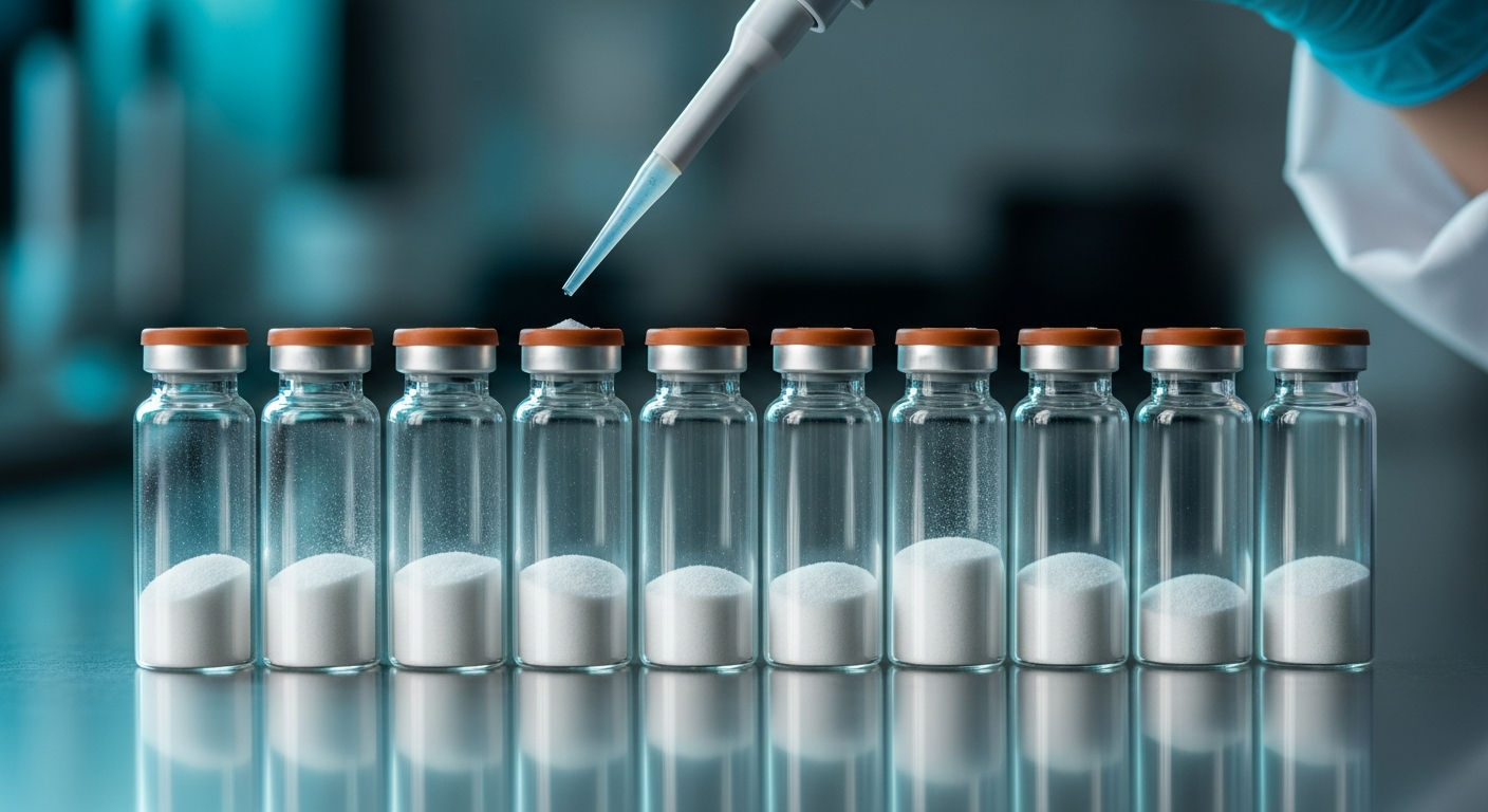
Proper preparation of the lyophilised reagent is paramount for maintaining molecular stability and ensuring assay reproducibility. Researchers must employ a sterile bacteriostatic reconstitution solution to dissolve the powder. This specific solvent prevents premature enzymatic degradation by inhibiting microbial protease activity and maintains the structural integrity of the dinucleotide during short-term cold storage.
When preparing stock solutions, investigators should gently swirl the vial rather than vortexing. Vigorous mechanical agitation can induce shear stress, potentially compromising the delicate N-glycosidic bonds. Once reconstituted, the solution requires immediate transfer to a controlled cold-storage environment, typically maintained at -80 degrees Celsius, to arrest spontaneous hydrolysis.
For comprehensive technical data regarding molecular weight, purity parameters, and specific storage guidelines, laboratory personnel should consult the Specification Sheet. This document outlines the exact physicochemical properties required for stringent in-vitro applications and provides essential safety data for laboratory risk assessments.
Advanced In-Vitro Assay Methodologies
Experimental designs often incorporate sophisticated fluorometric assays to monitor sirtuin activity continuously. By introducing a synthetic peptide substrate tagged with a specific fluorophore, researchers can measure the fluorescence emitted upon deacetylation. The addition of exogenous coenzyme initiates the reaction, allowing for real-time kinetic tracking and the generation of highly accurate progress curves.
High-resolution immunoblotting techniques offer another robust method for analysing downstream targets and confirming sirtuin activity. Following the controlled incubation of cell lysates with the exogenous coenzyme, investigators can utilise specific antibodies to quantify the acetylation status of structural proteins or transcription factors, such as p53 or PGC-1alpha. These protocols require precise timing and the rapid addition of denaturing buffers to accurately capture transient biochemical states before spontaneous degradation occurs.
Liquid chromatography-electrospray ionisation tandem mass spectrometry (LC-MS/MS) serves as the gold standard for quantifying the precise ratio of oxidised to reduced forms within cellular extracts. This advanced analytical technique separates the metabolites based on their distinct retention times and mass-to-charge ratios. Accurate measurement of this ratio is crucial for evaluating the overall redox environment of the experimental model.
Stability and Degradation Kinetics
The half-life of the reconstituted coenzyme in an aqueous environment is highly dependent on ambient pH and thermodynamic variables. Alkaline conditions rapidly accelerate the base-catalysed hydrolysis of the N-glycosidic bond, yielding biologically inactive byproducts. Therefore, researchers must carefully buffer the sterile bacteriostatic reconstitution solution to a neutral or slightly acidic pH, typically between 6.0 and 7.0, to maximise shelf life, synthesise stable aliquots, and preserve enzymatic reactivity.
Aliquoting the primary stock solution into single-use microcentrifuge tubes minimises detrimental freeze-thaw cycles. Repeated temperature fluctuations induce structural shearing and significantly reduce the concentration of the active coenzyme. Implementing strict inventory management protocols and utilising dedicated cold-storage units ensures consistent reagent quality across multiple, independent experimental runs.
The primary degradation products, including nicotinamide and cyclic ADP-ribose, can act as potent non-competitive inhibitors of sirtuin enzymes. Accumulation of these byproducts in the assay buffer will artificially suppress the measured catalytic rate. Consequently, researchers must prepare fresh working dilutions immediately prior to initiating the enzymatic reaction to prevent data artifact generation.
To source high-purity reagents for these highly sensitive assays, investigators can explore the catalogue at Amino Peptides. Procuring analytically validated compounds is the absolute first step in establishing a reliable, reproducible experimental baseline for complex metabolic research.
Kinetic Modelling and Structural Biology
Mathematical modelling of sirtuin kinetics requires precise, verifiable initial concentration data. Researchers plot the initial velocity of the deacetylation reaction against varying concentrations of the coenzyme to generate saturation curves. This classical Michaelis-Menten approach yields the specific Km and Vmax values, defining the fundamental catalytic efficiency of the specific sirtuin isoform under investigation.
Competitive inhibition assays further characterise the architecture of the binding pocket. By introducing structural analogues, scientists can map the specific amino acid residues involved in substrate recognition. These advanced structural biology techniques rely on X-ray crystallography and nuclear magnetic resonance spectroscopy to visualise the enzyme-substrate complex at atomic resolution.
The integration of CRISPR-Cas9 genetic editing technology allows for the selective knockout of specific sirtuin genes in immortalised cell lines. Subsequent introduction of the coenzyme isolates the metabolic impact of the remaining active isoforms. This reductionist approach is absolutely vital for untangling the complex, interconnected web of NAD+-dependent cellular signalling pathways.
Subcellular compartmentalisation plays a critical role in regulating these biochemical reactions. Distinct pools exist within the cytosol, mitochondria, and nucleus. In-vitro assays must frequently employ isolated organelle fractions to accurately assess the localised activity of specific sirtuins, such as the mitochondrial SIRT3 or the nuclear SIRT6, ensuring spatial dynamics are properly represented.
Frequently Asked Questions (In-Vitro Research)
What is the optimal pH for storing reconstituted NAD+ solutions?
The structural integrity of the dinucleotide is best preserved in a slightly acidic to neutral environment, strictly maintained between pH 6.0 and 7.0. Alkaline conditions rapidly induce the base-catalysed hydrolysis of the nicotinamide-ribose bond, rendering the molecule biologically inactive for sirtuin assays.
How does the choice of reconstitution solvent affect sirtuin assay reproducibility?
Employing a sterile bacteriostatic reconstitution solution prevents microbial contamination and inhibits exogenous protease activity. Pure deionised water lacks the necessary buffering capacity and antimicrobial properties, leading to rapid spontaneous degradation of the coenzyme and highly variable kinetic data across independent assay replicates.
Can fluorometric assays distinguish between SIRT1 and SIRT2 activity in-vitro?
While both specific enzymes require the same dinucleotide coenzyme for activation, methodological specificity is achieved by selecting distinct acetylated peptide substrates. SIRT1 and SIRT2 exhibit significantly different binding affinities for specific amino acid sequences flanking the target acetyl-lysine residue, allowing researchers to isolate isoform-specific activity.
Scientific Bibliography
- Imai, S., & Guarente, L. (2014). NAD+ and sirtuins in aging and disease. Trends in cell biology, 24(8), 464-471. View source
- Cantó, C., Menzies, K. J., & Auwerx, J. (2015). NAD+ metabolism and the control of energy homeostasis: a balancing act between mitochondria and the nucleus. Cell metabolism, 22(1), 31-53. View source
- Verdin, E. (2015). NAD⁺ in aging, metabolism, and neurodegeneration. Science, 350(6265), 1208-1213. View source
- Sauve, A. A., Wolberger, C., Schramm, V. L., & Boeke, J. D. (2006). The biochemistry of sirtuins. Annual review of biochemistry, 75, 435-465. View source
- Avalos, J. L., Celic, I., Muhammad, S., Cosgrove, M. S., Boeke, J. D., & Wolberger, C. (2002). Structure of a Sir2 enzyme bound to an acetylated p53 peptide. Molecular cell, 10(3), 523-535. View source
- Zhao, K., Harshaw, R., Chai, X., & Marmorstein, R. (2004). Structural basis for nicotinamide cleavage and ADP-ribose transfer by NAD+-dependent Sir2 histone/protein deacetylases. Proceedings of the National Academy of Sciences, 101(23), 8563-8568. View source
- Denu, J. M. (2005). The Sir2 family of protein deacetylases. Current opinion in chemical biology, 9(5), 431-440. View source