
Optimising Assay Conditions for the Quantification of Tesamorelin-Mediated cAMP Accumulation in Cultured Pituitary Cells
30th Jul 2026

Measuring cyclic adenosine monophosphate (cAMP) inside a cell is the standard way to test how well growth hormone-releasing hormone (GHRH) analogues work in a laboratory setting. Tesamorelin is a synthetic version of GHRH. It has an extra trans-3-hexenoic acid group attached to its structure, which stops enzymes from breaking it down as quickly as natural GHRH in test tubes. When this molecule attaches to the GHRH receptor on a cell surface, it forces the receptor to change shape. This change signals a G-protein inside the cell to swap a molecule called GDP for GTP, activating the system. To measure this reaction correctly, researchers must set exact conditions for the cell cultures. This means testing the exact number of cells, timing the exposure, and choosing the right liquid mixtures to track receptor activation. Scientists looking for high-purity materials for these tests can access specific compounds via our peptide research portal.
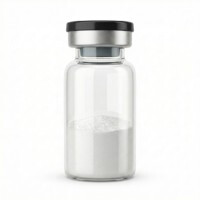
Tesamorelin (GHRH Analogue) |,, &
HPLC-verified lyophilised compound, UK-domestic supply. Manufactured under controlled conditions for qualified in-vitro laboratory research.
View Reagent Profile ›Choosing the right type of cell is the first step for a reliable cAMP test. Primary rat pituitary cells act most like cells in a living animal, but they are difficult to use. Every batch is different, and the mixture of cell types creates inconsistent data. To solve this problem, laboratories use immortal cell lines that divide continuously. The GH3 rat somatotroph cell line works well because it naturally produces GHRH receptors and can manufacture growth hormone in vitro. Another option is using standard laboratory cells, such as HEK-293 or CHO lines, modified to carry the human GHRH receptor. These modified cells provide a uniform test group with very high receptor numbers, making the final signal much easier to read against the background noise.
No matter which cell model researchers choose, they must prepare the cells carefully. The cells require a period of serum starvation before the test begins. This means removing the nutrient-rich blood serum from their liquid food. The serum contains hidden factors that can constantly trigger the cells and raise their resting cAMP levels. Soaking the cells for four to six hours in a simple liquid containing 0.1% bovine serum albumin (BSA) settles the cells down. This step stabilises the starting cAMP levels without killing the cells. It removes background noise from outside growth factors that might accidentally activate other internal pathways and ruin the tesamorelin data.
Key Takeaways for Assay Optimisation
- Serum Starvation: Starving the cells of serum for four to six hours lowers resting cAMP levels and widens the measurable range of the test.
- Phosphodiesterase Inhibition: The test mixture must include a blocking chemical, such as IBMX, to stop enzymes from destroying the new cAMP immediately.
- Incubation Kinetics: Technicians must stop the test at exact times, usually at 30 minutes. This catches the highest cAMP point before the receptors shut down and hide inside the cell.
- Buffer Composition: The test liquid needs natural levels of magnesium ions (Mg2+). Magnesium acts as a necessary partner for the enzyme that actually builds the cAMP.
Inside a living cell, cAMP does not last long. Natural enzymes called phosphodiesterases (PDEs) constantly break it down into an inactive molecule called 5'-AMP. To build up enough cAMP to measure after adding the tesamorelin reagent, researchers must temporarily block these enzymes. They do this by adding a chemical blocker called IBMX to the test liquid. The laboratory must test different amounts of IBMX to find the perfect dose. While 0.5 mM is standard, some cell types need more or less depending on how many PDE enzymes they produce. If there is not enough blocker in the liquid, the cAMP disappears too quickly. This missing signal makes the tesamorelin look weaker than it really is during laboratory analysis.

The test liquid must keep a natural pH of 7.4 and the correct salt balance. Researchers prefer a mixture called Hanks' Balanced Salt Solution (HBSS) with HEPES buffer. HEPES keeps the pH stable when the cells sit on a workbench outside their incubator. Next, the liquid must contain magnesium. The magnesium ions lock into the cell's cAMP-building engine, helping it break apart ATP molecules to form new cAMP. Finally, adding a simple protein called BSA stops the sticky peptide from clinging to the sides of the plastic test tubes.
Older methods for measuring cAMP used radiation or complex enzyme tests. These older tests required technicians to wash the cells multiple times, which added errors and took hours to finish. Today, major screening laboratories use simple mix-and-read tools. A method called Homogeneous Time-Resolved Fluorescence (HTRF) is the most common choice for tracking cAMP inside cells. This test uses two glowing tags: one attached to an antibody and another attached to a fake cAMP molecule. When the cell is resting, the two glowing tags link together and shine brightly at a specific light wavelength (665 nm). When the cell reacts to tesamorelin and makes real cAMP, the real cAMP pushes the fake glowing molecule out of the way. This breaks the link between the tags, and the bright 665 nm light drops.
HTRF is very sensitive, and it rarely gets confused by cloudy liquids or coloured chemicals in the test plates. Researchers looking at how different compounds trigger chemical pathways in cell cultures can also test immunomodulatory peptide categories. Testing different peptide classes reveals exactly how different cell receptors change internal cAMP levels under a microscope.
A standard laboratory protocol for the cAMP test follows these strict steps. First, place 15,000 cells into each well of a white 96-well microplate using 100 µL of standard growth liquid. Leave the plates in a 37°C incubator with 5% CO2 for 24 hours. Second, remove the liquid, rinse the cells with warm salt water, and add a simple liquid without serum. Leave them for five hours to starve the cells of outside signals. Third, mix ten different strengths of the peptide in a test buffer containing the IBMX blocker. Fourth, remove the starvation liquid and add 20 µL of the peptide mixtures to the cells. Let the plate sit for exactly 30 minutes at 37°C. Fifth, stop the reaction by adding the glowing HTRF tags and chemical soap to break the cells open. Leave the plate in the dark at room temperature for 60 minutes. Sixth, put the plate into a machine that reads the glowing light at 620 nm and 665 nm. The machine uses a basic maths formula to cancel out any small optical errors in the plastic.
The raw light readings from the machine do not directly tell the researcher the cAMP amounts. Technicians must convert the light ratios into true cAMP numbers using a standard reference curve built during the same test run. Once the laboratory calculates the absolute cAMP levels, usually measured in nanomoles, they plot these points on a graph against the different doses of the peptide.
The graphing software uses a mathematical formula called a four-parameter logistic (4PL) equation to draw a curve through the data points. This formula tracks four specific details. It finds the lowest baseline signal, the highest peak signal, the slope of the line, and the EC50 value. The EC50 shows the exact peptide dose needed to hit half of the maximum cell response. A clean, successful laboratory test will produce a smooth, S-shaped curve on the graph, confirming the cell response is genuine and measurable.
Frequently Asked Questions
1. Why is serum starvation essential prior to stimulating cells with GHRH analogues?
Animal serum contains random hormones and fats that constantly trigger the cell's internal receptors. This background noise makes the resting cAMP levels jump up and down, which shrinks the measurable window of the test. Starving the cells of this serum resets their resting state. This step guarantees that any new cAMP tracked in the test comes strictly from the added peptide and not from random food signals.
2. What are the consequences of omitting a phosphodiesterase inhibitor from the stimulation buffer?
Leaving out a blocking chemical like IBMX lets the cell's natural enzymes destroy the new cAMP almost immediately. If the cell destroys cAMP faster than the test peptide can force the cell to build it, the total cAMP levels will barely rise. The test results will look completely flat. This makes it impossible to see if the peptide actually worked, and it makes the compound appear much weaker than it actually is in vitro.
3. How does cell seeding density influence the calculated EC50 of GHRH analogues?
The number of cells in the dish determines the ratio of peptide molecules to cell receptors. If a technician packs too many cells into a single well, the cells absorb all the available peptide too quickly. This skews the graph and makes the chemical look less effective. Laboratories must test different cell amounts to find a perfect, single layer of cells, usually between 10,000 and 20,000 per well, to get accurate reading values.
Scientific References
- Mayo, K. E., Miller, T. L., DeAlmeida, V., et al. (2003). The growth hormone-releasing hormone receptor: signal transduction, gene expression, and physiological regulation in vitro. Annals of the New York Academy of Sciences, 992(1), 212-221. View published research
- Ferdinand, R., Besser, G. M., & Trainer, P. J. (2007). Structural modifications and enzymatic stability of growth hormone-releasing hormone analogues in pituitary cell cultures. Journal of Endocrinology, 194(2), 321-329. View published research
- Degorce, F., Card, A., Soh, S., et al. (2009). HTRF ligand binding and cell-based cAMP assays for G-protein coupled receptors. Current Chemical Genomics, 3, 22-32. View published research
- Billestrup, N., Swanson, L. W., & Vale, W. (1986). Growth hormone-releasing factor stimulates somatotroph proliferation in primary anterior pituitary cultures. Proceedings of the National Academy of Sciences, 83(11), 3751-3755. View published research
⚠️ Research Use Only Disclaimer: All peptides and compounds are sold strictly for in-vitro laboratory research purposes only. Not intended for human or veterinary use, not a dietary supplement, and not approved to diagnose, treat, cure, or prevent any disease or condition. For use by qualified researchers in suitably equipped laboratory environments only. Amino Peptides Ltd operates in full compliance with UK MHRA regulations and applicable EU research chemical guidelines.