
Optimising Tesamorelin Reconstitution Protocols for Long-Term In-Vitro Stability
11th Sep 2026

What You Need to Know About Tesamorelin
- Tesamorelin is a 44-residue GHRH analogue with a trans-3-hexenoic modification on the N-terminus.
- Published models follow GHRH-receptor signalling and later metabolic readouts in the assay.
- Readers compare it with Sermorelin, which is the shorter 29-residue GHRH fragment.
- The longer sequence is only meaningful if the vial is 99%+ HPLC-verified and stored at 2°C–8°C.
How Tesamorelin Works Inside Laboratory Cell Models
Tesamorelin is a synthetic forty-four-amino-acid analogue of growth hormone-releasing factor that carries a protective trans-3-hexenoic acid group at its N-terminus. In laboratory models, this modified cap shields the peptide from rapid enzymatic breakdown so it can bind pituitary receptors and support sustained cellular energy and tissue remodelling signals.
To understand why optimising Tesamorelin reconstitution protocols for long-term in-vitro stability matters so much, it helps to look at how the molecule is built. Natural growth hormone-releasing hormone has a very short lifespan in solution and in biological fluids because an enzyme called dipeptidyl peptidase-4 snips the chain between the second and third amino acids. By attaching a six-carbon hexenoyl group to the tyrosine residue at position one, chemists gave Tesamorelin a sturdy molecular helmet. This structural change keeps the sequence intact long enough to lock onto growth hormone-releasing hormone receptors on somatotroph cells.
Once bound in a petri dish or preclinical model, the receptor activates an internal messenger system driven by cyclic adenosine monophosphate, commonly known as cAMP. Think of cAMP as a relay runner that wakes up protein kinase A and prompts the cell to release stored growth hormone in natural-looking pulses rather than a flat, continuous wave. Those pulses then travel to liver cells in experimental models to encourage the production of insulin-like growth factor-1, a key driver of cellular energy, lipid oxidation, and structural tissue recovery.
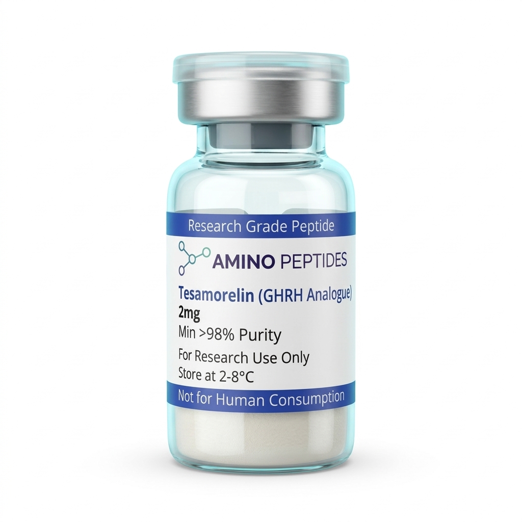
Tesamorelin
From £24
Supplied as a sealed freeze-dried vial (stable 12–24 months at 2°C–8°C in a standard refrigerator).
Shop Tesamorelin →However, a forty-four-amino-acid chain is physically large compared to shorter fragments, and it folds into an alpha-helical shape when it interacts with water and lipid membranes. If a researcher mixes the lyophilised powder too roughly, uses the wrong liquid carrier, or leaves the liquid warm on a benchtop, those hydrophobic sections can clump together into inactive beta-sheet fibrils. Once a peptide forms these sticky aggregates or suffers oxidation at its methionine residue, it can no longer fit neatly into the receptor pocket. Gentle mixing along the glass wall of the vial and choosing the right solvent pH keep the alpha-helix free to do its job in the dish.
Published studies also look at how this receptor signalling influences adipocyte cultures and mitochondrial turnover. When the peptide remains intact in solution, researchers observe steady activation of lipolytic enzymes that break down stored triglycerides without disturbing baseline glucose balance in the surrounding media. Every one of these clean, repeatable observations depends on keeping the full forty-four-amino-acid chain in its native, unfolded, and unoxidised state from the moment the liquid touches the freeze-dried cake.
What Published Studies Observe Across Different Timelines
When investigators track Tesamorelin in laboratory models, the observed outcomes split clearly into fast chemical signalling and slower structural changes. Because optimising Tesamorelin reconstitution protocols for long-term in-vitro stability often means using the same stock solution across several weeks of testing, knowing what happens at each stage helps researchers spot whether their reconstituted batch is still performing at full strength.
During the first five to thirty minutes of in-vitro exposure, the action is purely electrical and chemical. In isolated pituitary cell cultures, adding a fresh measured level of Tesamorelin triggers an immediate spike in intracellular cAMP and calcium ion movement across the cell membrane. Within one to four hours, researchers can measure a clear, pulsatile release of growth hormone into the surrounding culture media. If a reconstituted vial has been sitting in a warm room or was mixed with an incompatible buffer, this early cAMP spike drops off noticeably, giving the lab an early warning that the peptide chain has begun to degrade or stick to the inside of the glass vial.
Moving into the twenty-four to seventy-two-hour window, the focus in preclinical models shifts toward secondary messenger transcription and liver cell signalling. At this stage, published studies document a steady rise in insulin-like growth factor-1 mRNA expression and early shifts in lipid metabolism enzymes. Cultured adipocytes begin showing increased hormone-sensitive lipase activity, which starts liberating fatty acids from internal lipid droplets to support cellular energy demands.
Reconstitution calculator
Choose a vial mass and a bacteriostatic reconstitution solution volume. The figure is the resulting concentration.
Concentration: 2.50 mg/mL
Per 0.01mL (10µL): 25.0 mcg
Open the full calculatorBacteriostatic reconstitution solutionOver multi-week laboratory protocols lasting two to eight weeks, the slower structural and metabolic changes become visible. In longer preclinical models, sustained pulsatile signalling leads to measurable reductions in visceral adipose tissue volume, improved mitochondrial density in skeletal muscle samples, and stronger collagen matrix organisation during tissue remodelling experiments. Researchers studying peripheral nerve recovery in animal models have also noted steadier axonal regeneration over four-to-six-week observation windows when the peptide concentration is kept consistent.
Running a four-week or six-week experiment requires a stock solution that behaves the exact same way on day twenty-eight as it did on day one. If deamidation at the asparagine residues or aggregation slowly lowers the active concentration inside the vial, the later structural readouts will look artificially weak. Splitting a newly mixed batch into smaller single-use aliquots and freezing them at minus twenty or minus eighty degrees Celsius is the simplest way to keep multi-week timelines consistent from start to finish.
Comparison of In-Vitro Reconstitution Solvents and Storage Conditions for Tesamorelin
| Reconstitution Medium | Target pH Range | Storage Temperature | Observed In-Vitro Stability | Primary Degradation Risk |
|---|---|---|---|---|
| Sterile Deionised Water (with 4 to 5% Mannitol) | 5.0 to 6.5 | 2 to 8 degrees Celsius | Up to 7 days with minimal structural loss | Gradual hydrolysis if left at room temperature |
| Bacteriostatic Reconstitution Solution (0.9% Benzyl Alcohol) | 5.5 to 6.5 | 2 to 8 degrees Celsius | 10 to 14 days under strict refrigeration | Alcohol-induced hydrophobic aggregation at higher concentrations |
| 0.9% Sodium Chloride (Normal Saline) | 5.0 to 7.0 | 2 to 8 degrees Celsius | Less than 24 hours (Not recommended) | Rapid salting-out effect and visible white precipitation |
| 10 mM Sodium Acetate Buffer | 4.5 to 5.5 | Minus 20 degrees Celsius (Single-use aliquots) | Up to 60 days when protected from light | Mechanical shear stress if subjected to repeated freeze-thaw cycles |
| Phosphate-Buffered Saline (PBS) | 7.2 to 7.4 | 20 to 25 degrees Celsius | Under 6 hours | Accelerated deamidation of asparagine residues at neutral pH |
Pairing Tesamorelin With Complementary Research Peptides
In laboratory research, scientists rarely look at a single signalling pathway in isolation. Because Tesamorelin works through the growth hormone-releasing hormone receptor, published studies frequently pair it with complementary compounds to observe how multiple cellular pathways interact during tissue recovery and metabolic regulation.
The most common pairing in preclinical receptor studies combines Tesamorelin with a growth hormone secretagogue receptor agonist such as Ipamorelin. While Tesamorelin steps on the accelerator by raising cAMP levels inside somatotroph cells, Ipamorelin mimics ghrelin at a completely separate receptor driven by phospholipase C and inositol trisphosphate. At the same time, Ipamorelin quiets down somatostatin, the natural brake pedal that normally slows hormone release. When both receptors are engaged in a laboratory model, the resulting pulse is much larger than either peptide produces on its own, allowing researchers to study maximal pulsatile output using lower study amounts of each reagent.
For investigations focused on cellular energy and fat oxidation, laboratories often explore Tesamorelin alongside MOTS-c and NAD+ in multi-variable cell culture designs. While Tesamorelin encourages the release of stored fatty acids from lipid droplets, mitochondrial peptides and coenzymes help the cell's powerhouses convert those freed fatty acids into usable adenosine triphosphate. Observing both stages together gives researchers a complete view of how mobilisation and oxidation work side by side in metabolically stressed cells.
In tissue remodelling and structural recovery models, Tesamorelin is also studied alongside the pentadecapeptide BPC-157 or Thymosin Beta-4 fragments. In these setups, Tesamorelin supports systemic growth factor signalling and collagen synthesis, while BPC-157 promotes local angiogenesis and fibroblast migration across the extracellular matrix. Together, they allow investigators to track how systemic anabolic signals and local microvascular repair complement one another in tendon, ligament, and muscle cultures.
One practical rule applies to all of these combinations: never mix different peptides inside the same storage vial for long-term refrigeration. Even when two sequences share a similar pH preference, placing a forty-four-amino-acid chain like Tesamorelin in the same bottle as a smaller peptide changes the ionic strength of the liquid and speeds up cross-aggregation. Optimising Tesamorelin reconstitution protocols for long-term in-vitro stability means keeping each compound in its own dedicated vial, stored at its own ideal pH, and only combining measured levels in the assay dish right when the experiment begins.
Why Purity, Solvents, and Cold Storage Protect Your Vial
Even the most careful benchtop technique cannot rescue a peptide that left the synthesiser full of chemical leftovers. When optimising Tesamorelin reconstitution protocols for long-term in-vitro stability, the purity of the freeze-dried cake and the exact makeup of the liquid solvent determine how long the peptide stays dissolved and active.
During solid-phase synthesis, chemists build Tesamorelin one amino acid at a time and use trifluoroacetic acid, known as TFA, to cleave the finished chain and strip away protective side groups. If a manufacturer skips thorough post-synthesis ion-exchange cleanup, residual TFA stays trapped in the lyophilised powder. In a long forty-four-amino-acid peptide, excess TFA lowers the pH uncontrollably once liquid is added, eats away at sensitive peptide bonds through acid hydrolysis, and proves toxic to living cells in culture. Choosing vials verified at ninety-nine percent or higher purity by High-Performance Liquid Chromatography, with TFA salts properly removed or exchanged for benign acetate counter-ions, prevents the powder from digesting itself in solution.
Solvent choice is the next critical piece of the puzzle. Tesamorelin has an isoelectric point near pH 8.5 and contains hydrophobic patches that dislike neutral, unbuffered water. If a researcher adds plain sterile water, the liquid can sit too close to a pH where the molecules lose their mutual electrostatic repulsion and clump into cloudy gel fibres. For standard multi-week refrigerated storage, a quality bacteriostatic reconstitution solution containing 0.9 percent benzyl alcohol keeps microbial growth at bay while providing a clean aqueous environment. When a specific assay calls for a higher concentration stock or shows slight cloudiness upon mixing, using diluted acetic acid gently lowers the pH away from the peptide's isoelectric point, giving the chains a strong positive charge so they repel each other and stay crystal clear in solution.
When adding your chosen liquid, angle the needle so the stream glides slowly down the inside glass wall rather than blasting the fragile lyophilised cake directly. Never shake the vial; shaking traps air bubbles and creates air-water interfaces that unfold the alpha-helix like egg whites beaten into foam. Instead, roll the vial gently between your palms until every speck dissolves.
Once mixed, temperature controls the speed of chemical wear and tear. Store the reconstituted vial upright in a dedicated laboratory refrigerator kept between two and eight degrees Celsius, away from light and door vibrations. For studies stretching past two or three weeks, divide the fresh solution into low-bind polypropylene micro-vials and freeze them once to prevent repeated freeze-thaw crystal damage.
Frequently Asked Questions
Why are researchers focused on optimising Tesamorelin reconstitution protocols for long-term in-vitro stability?
Researchers search for ways to stabilise Tesamorelin because its forty-four amino acid chain breaks down rapidly once mixed into a liquid. Optimising the reconstitution protocol with a bacteriostatic reconstitution solution prevents premature degradation, allowing laboratory models to yield accurate, repeatable data on growth hormone-releasing factor signalling and tissue remodelling.
What is the typical timeline for observing Tesamorelin activity in laboratory models?
Published studies show that pituitary cell receptor signalling begins within minutes of introducing a measured level of reconstituted Tesamorelin in vitro. However, broader biological shifts in preclinical models, such as measurable changes in lipid metabolism and cellular recovery markers, generally require continuous observation across a four to twelve-week study timeline.
Can Tesamorelin be combined with other peptides during in-vitro research?
Yes, researchers frequently pair Tesamorelin with complementary growth hormone secretagogues like Ipamorelin in laboratory models to analyse synergistic receptor signalling. However, to maintain long-term in-vitro stability, each compound should be mixed in its own vial using a separate bacteriostatic reconstitution solution rather than combining them into a single storage container.
How do purity and fridge storage impact reconstituted Tesamorelin stability?
High initial purity of at least ninety-nine percent and strict fridge storage between two and eight degrees Celsius are essential for preserving reconstituted Tesamorelin. Keeping the vial cold and shielded from light slows down hydrolysis and oxidation, which maintains the exact protocol concentration needed for reliable multi-week laboratory experiments.
Bibliography
- Ferdinandi ES, Brazeau P, High K, et al. Non-preclinical pharmacology and safety evaluation of TH9507, a human growth hormone-releasing factor analogue. Basic & preclinical Pharmacology & Toxicology. 2007;100(1):49-58. View published research
- Manning MC, Chou DK, Murphy BM, Payne RW, Katayama DS. Stability of protein pharmaceuticals: an update. Pharmaceutical Research. 2010;27(4):544-575. View published research
- Wang W. Instability, stabilization, and formulation of liquid protein pharmaceuticals. International Journal of Pharmaceutics. 1999;185(2):129-188. View published research
- Zapadka KL, Becher FJ, Gomes Dos Santos AL, Jackson SE. Factors affecting the physical stability (aggregation) of peptide signalling. Interface Focus. 2017;7(6):20170030. View published research
⚠️ Research Use Only Disclaimer: All peptides and compounds are sold strictly for in-vitro laboratory research purposes only. Not intended for human or veterinary use, not a dietary supplement, and not approved to diagnose, treat, cure, or prevent any disease or condition. For use by qualified researchers in suitably equipped laboratory environments only. Amino Peptides Ltd operates in full compliance with UK MHRA regulations and applicable EU research chemical guidelines.