
Quantifying Results: Analytical Methods for Measuring Peptide Degradation
4th Jul 2026
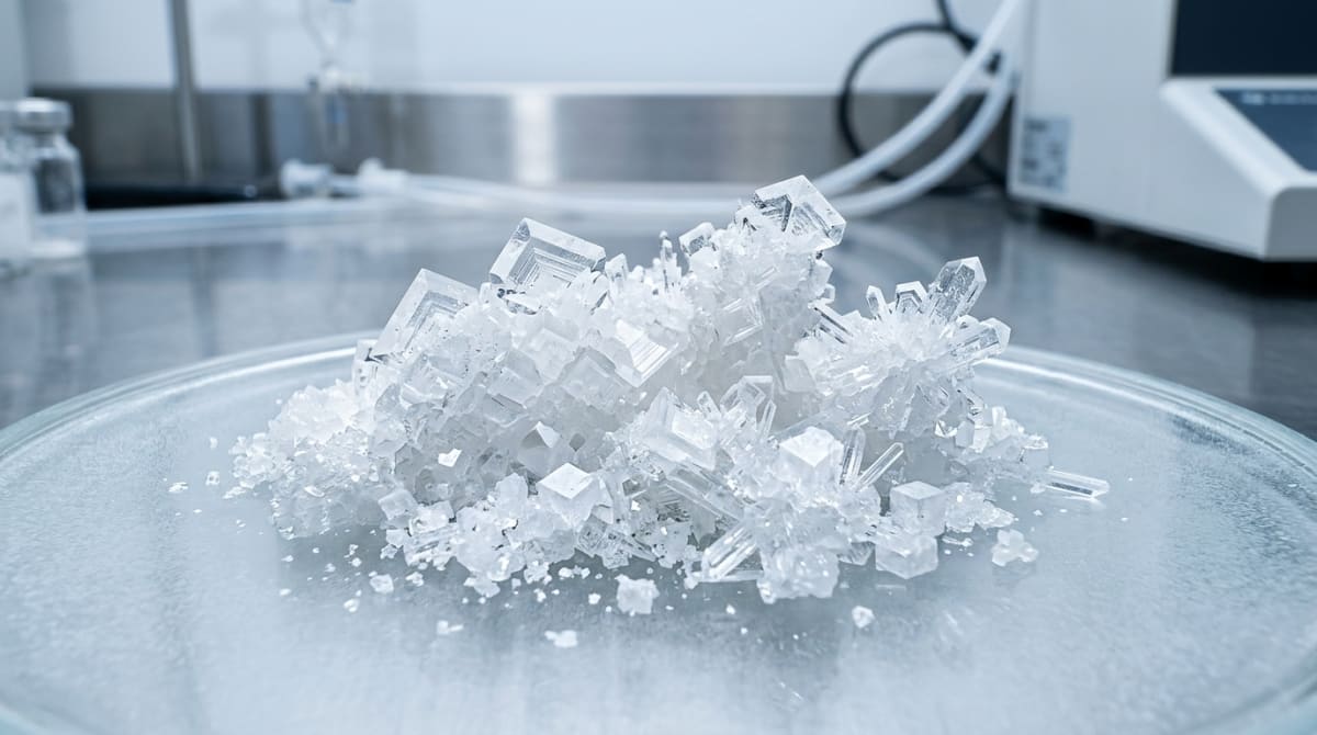
Synthetic peptide reagents require precise quality control during in-vitro investigations. Built from amino acids joined by amide bonds, these molecular chains frequently experience chemical and physical degradation over time. Quantifying structural shifts ensures baseline experimental validity. Analytical chemists depend on specific instrumentation to monitor molecular stability, verifying that peptide structures remain intact across cellular assays. This review outlines primary analytical techniques for measuring degradation rates, supporting baseline reagent integrity.
Mapping degradation pathways establishes baseline compound stability. Polypeptides undergo specific chemical modifications upon exposure to environmental variables, including temperature fluctuations, pH changes, and ultraviolet light. Identifying these breakdown mechanisms aids researchers in sourcing materials from a reputable UK peptide supplier, confirming pre-assay stability.
The primary chemical degradation pathways include:
- Deamidation: A hydrolytic reaction where side-chain amide groups in glutamine or asparagine residues convert to carboxylic acids. This alters the net molecular charge and physical behaviour.
- Oxidation: Primarily affecting methionine, cysteine, and tryptophan residues. Exposure to ambient oxygen or reactive oxygen species generates sulfoxides, modifying peptide hydrophobicity.
- Hydrolysis: The cleavage of peptide bonds, which yields smaller fragments. This reaction accelerates under extreme pH levels or elevated temperatures in aqueous environments.
- Isomerisation: The conversion of L-aspartyl residues into D-aspartyl forms, changing spatial configuration without shifting molecular weight.
- Disulphide Exchange: The rearrangement of disulphide bonds, disrupting the tertiary structure of folded complex peptides.
Surrounding medium pH dictates both the rate and mechanism of chemical degradation. For example, asparagine deamidation accelerates in neutral to alkaline buffers (pH 7 to 9) through a succinimide intermediate. Highly acidic environments (pH 1 to 2) drive direct side-chain amide hydrolysis. This acid-catalysed reaction proceeds slowly but increases the risk of peptide backbone cleavage. Characterising these pH-dependent variables allows investigators to specify precise buffer systems for in-vitro protocols.

Figure 1: A clean, abstract 2D node-and-network biological vector graphic on a laboratory interface screen, showcasing molecular connections without text.
Reversed-Phase High-Performance Liquid Chromatography (RP-HPLC) provides the standard methodology for separating intact peptides from degradation fragments. The technique depends on hydrophobic interactions between the analyte, the mobile phase, and the stationary phase of the column. Configurations include:
- Stationary Phase: Silica-based columns bonded with alkyl chains, typically C18 or C8, supplying the necessary hydrophobic surface.
- Mobile Phase: A gradient system consisting of an organic solvent, usually acetonitrile, and water, supplemented with an ion-pairing agent like trifluoroacetic acid (TFA) to sharpen peak profiles.
- Detection: Ultraviolet (UV) absorbance detection at 210 nm or 214 nm, corresponding to the peptide bond absorption wavelength, enabling sensitive identification of all species.
Modified fragments present distinct retention times compared with the parent molecule. Deamidated products express higher polarity, typically eluting earlier, whereas oxidised variants display shifted retention profiles dictated by the specific altered residue. Analysts calculate absolute purity percentages by comparing the intact peptide peak area against total integrated peak areas. Wide-pore columns (300 Angstroms) prevent steric hindrance during polypeptide separation. Maintaining column temperatures between 30 and 40 degrees Celsius ensures reproducible retention, while a shallow, linear mobile phase gradient separates closely eluting degradants.
While RP-HPLC yields quantitative resolution based on peak area, Liquid Chromatography-Mass Spectrometry (LC-MS) provides precise structural identification. Coupling chromatographic separation with mass analysis permits investigators to assign exact molecular weights to isolated peaks, confirming degradant identity through:
- Electrospray Ionisation (ESI): A soft ionisation technique preserving delicate peptide structures during transition into the gas phase, permitting accurate mass determination of intact molecules.
- Tandem MS (MS/MS): This approach sequences fragment ions, pinpointing the exact amino acid residue that has undergone modification, such as locating specific oxidised methionine residues.
Structural characterisation confirms whether a secondary peak indicates a minor oxidation event or a severe sequence cleavage capable of skewing in-vitro data. High-resolution mass spectrometry (HRMS) platforms deliver sub-parts-per-million mass accuracy. Such precision separates modifications presenting similar mass shifts. Furthermore, tandem MS sequences the resulting fragments, mapping the exact modification site along the carbon backbone.
Secondary analytical techniques deliver additional data to construct a comprehensive stability profile:
- Capillary Electrophoresis (CE): Separates molecules via charge-to-size ratios within an electric field. This approach identifies charge variants resulting from deamidation, supplying an alternative separation mechanism to hydrophobic chromatography.
- Size Exclusion Chromatography (SEC): Necessary for detecting physical degradation, including aggregation or oligomerisation. Because large aggregates can elute in the void volume or bind irreversibly to RP-HPLC columns, SEC quantifies these high-molecular-weight species.
Dynamic Light Scattering (DLS) quantifies the hydrodynamic radius of suspended particles, identifying sub-micron aggregates before they become visible. This metric is highly relevant for larger polypeptides prone to self-association, as aggregate accumulation directly alters molecular kinetics during laboratory assays.
Quantification depends on the area-under-the-curve (AUC) method, calculating the relative peak area of the intact peptide against all detected impurities to generate a percentage purity value. Regulatory documentation requires validated analytical methods capable of differentiating active compounds from closely related degradants. This separation confirms that in-vitro data stems from stable, chemically defined entities, as noted in discussions on regulatory standards.
In-Vitro Analytical FAQ
What is peptide degradation?
Peptide degradation defines the chemical or physical alteration of a primary, secondary, or tertiary structure over time. In laboratory environments, this breakdown depletes the active peptide concentration, compromising in-vitro experiment validity. Chemical degradation encompasses covalent bond disruption or formation (hydrolysis, deamidation, oxidation). Physical degradation involves spatial conformational shifts, such as precipitation, aggregation, or surface adsorption.
What are peptide degradation products?
These products are molecular fragments, modified chains, or aggregated complexes generated during parent peptide breakdown. Examples include deamidated sequences, sulfoxide-bearing oxidised variants, hydrolytically cleaved fragments, and high-molecular-weight oligomers. Quantifying these byproducts is imperative, as their presence frequently disrupts receptor-binding assays or shifts solution thermodynamics, causing erratic assay readouts.
How does the degradation of polypeptides occur in a laboratory setting?
Degradation stems from environmental stress during storage and active handling. Elevated temperatures, ultraviolet light exposure (photolysis), extreme solvent pH, and mechanical shear forces accelerate the breakdown rate. Storing polypeptides in room-temperature aqueous buffers induces rapid hydrolysis and deamidation. Conversely, securing them as lyophilised powders at -20 degrees Celsius or below effectively arrests these degradative mechanisms.
Securing the integrity of in-vitro research demands strict, systematic quantification of peptide stability. Integrating high-resolution RP-HPLC with the molecular identification power of LC-MS allows laboratories to monitor degradation pathways and isolate impurities. Strict adherence to validated storage and reconstitution parameters guarantees that experimental reagents remain stable, producing reproducible laboratory data.
- D'Hondt, M., et al. (2014). Analytical quality-by-design approach for chimeric peptide stability-indicating HPLC method development. Journal of Pharmaceutical and Biomedical Analysis, 90, 108-116. View published research
- Ghafourian, T., et al. (2007). The chemical stability of monomeric and polymeric peptide formulations. Journal of Peptide Science, 13(12), 793-801. View published research
- Manning, M. C., et al. (2010). Stability of protein pharmaceuticals: an update. Pharmaceutical Research, 27(4), 544-575. View published research
⚠️ Research Use Only Disclaimer: All peptides and compounds are sold strictly for in-vitro laboratory research purposes only. Not intended for human or veterinary use, not a dietary supplement, and not approved to diagnose, treat, cure, or prevent any disease or condition. For use by qualified researchers in suitably equipped laboratory environments only. Amino Peptides Ltd operates in full compliance with UK MHRA regulations and applicable EU research chemical guidelines.