
Quantitative Analysis of Peptide-Membrane Interactions Using Cell-Free Fluorescence Assays
9th Oct 2026
What You Need to Know About Quantitative Analysis of Peptide-Membrane Interactions Using Cell-Free Fluorescence Assays
- Researchers search for these assays to measure how strongly peptides bind to cell borders without the messy background noise of whole living cells.
- The method works by shining light on glowing markers that shift colour or brightness the exact moment a peptide touches a synthetic fat bubble.
- Published studies observe that tracking these light shifts lets scientists calculate exact binding strengths, pore sizes, and lipid mixing speeds.
- Laboratory models show that tuning the electric charge of the artificial membrane completely changes how deeply a peptide sinks into the lipid layer.
How Glowing Dyes Track Cellular Energy and Membrane Repair
Cell-free fluorescence assays measure exactly how peptides bind to, bend, or cross synthetic cell barriers by tracking shifts in light emission. This method gives researchers a clear, numbers-based look at how specific amino acid chains support cellular energy and structural recovery without background noise from whole cells.
Every cell in the body relies on fatty outer walls and inner mitochondrial membranes to stay alive and make energy. When scientists study tissue repair research, they first need to know how a peptide physically touches these lipid layers. Whole living cells are busy and messy, making it hard to see single interactions. Instead, lab teams build simple, artificial lipid bubbles called liposomes or giant unilamellar vesicles. These synthetic bubbles mimic real biological walls, from skin surface barriers to energy-producing inner membranes.
To watch the interaction, scientists attach or mix in special glowing dyes. Some peptides naturally glow under ultraviolet light because they contain the amino acid tryptophan. When tryptophan moves from watery liquid into the fatty centre of a membrane, its light shifts to a shorter wavelength and shines brighter. Other tests trap glowing dye inside the lipid bubble. If a peptide loosens the lipid packing or opens a temporary pore, the dye leaks out and lights up the surrounding liquid. By measuring the exact brightness on a spectrofluorometer, investigators calculate binding strength, surface partitioning, and lipid stability. This shows how well a sequence can stabilise stressed mitochondrial walls or support outer membrane recovery in preclinical models.
This page is not tied to one stocked vial. Start with the bundles hub, BPC-157, or GHK-Cu — each is in stock for next-day UK dispatch.
Browse Full UK Peptide Catalogue →From Millisecond Lipid Binding to Long-Term Structural Shifts
Tracking a peptide inside a cell-free fluorescence setup happens across two very different clock speeds: immediate contact and slower structural settling. During the first phase, which takes milliseconds to a few seconds, electrostatic attraction pulls the charged amino acid chain toward the oppositely charged lipid heads. Stopped-flow fluorescence instruments capture this rapid flash of light as the sequence touches the water-lipid border. In published studies, tryptophan blue-shifts or Laurdan dye readings change almost instantly, showing that initial surface docking happens right after mixing the measured level of peptide into the buffer.
The second phase unfolds over several minutes to a few hours. Once the peptide sits on the outer layer, it begins to fold—often twisting into a neat spiral called an alpha-helix—and sinks deeper into the fatty core. During this window, scientists watch slower changes in fluorescence anisotropy, a measurement that tells them how stiff or fluid the membrane has become. If the protocol studies dye leakage or lipid mixing, the glowing signal climbs steadily for fifteen to sixty minutes before hitting a flat plateau. By comparing the fast first-second flash against the one-hour plateau, lab teams separate quick surface sticking from lasting physical remodelling of the lipid bilayer. This timeline shows whether a sequence simply coats the outside of a vesicle or permanently reorganises the lipid packing to support barrier recovery.
Common Cell-Free Fluorescence Assays for Peptide-Membrane Analysis
| Assay Method | Fluorescent Probe | Membrane Model | Primary Measurement |
|---|---|---|---|
| Tryptophan Blue-Shift Assay | Intrinsic tryptophan amino acid | Large Unilamellar Vesicles (LUVs) | Partition coefficient and burial depth in the lipid core |
| Calcein Leakage Assay | Self-quenching calcein dye | Synthetic lipid liposomes | Membrane permeabilisation and pore formation speed |
| Acrylamide Quenching | Tryptophan plus water-soluble quencher | Small Unilamellar Vesicles (SUVs) | Solvent exposure and peptide orientation at the surface |
| Laurdan Generalised Polarisation | Environment-sensitive Laurdan dye | Giant Unilamellar Vesicles (GUVs) | Lipid packing order and water penetration into the bilayer |
| FRET Lipid Mixing Assay | NBD and Rhodamine headgroup pair | Labelled and unlabelled LUV mixtures | Vesicle fusion and membrane stalk formation rates |
Pairing Membrane-Active Sequences in Combined Lab Protocols
In laboratory models, researchers rarely look at a single molecule in isolation when they can test how complementary sequences behave together on the same lipid surface. Quantitative fluorescence assays make it easy to run dual-peptide protocols and watch how two different chains share space on a synthetic membrane. One common pairing combines a surface-anchoring sequence like SS-31 (Elamipretide), which seeks out cardiolipin fats found in energy-producing mitochondrial walls, with a broader structural remodelling sequence such as TB-500 or BPC-157. By tagging each peptide with a different coloured fluorophore, scientists use a method called Förster Resonance Energy Transfer (FRET) to measure the exact distance between the two molecules on the lipid bubble.
When two compatible sequences sit within a few nanometres of each other, energy jumps from the first dye to the second, creating a distinct glow that only appears when they cooperate. Published studies show that an initial anchoring peptide can slightly change the curvature or surface charge of a lipid vesicle, making it much easier for a secondary recovery peptide to dock at a lower concentration. Conversely, testing combinations in a cell-free setup also reveals when two sequences crowd each other out and compete for the same lipid binding spots. Running these paired assays side by side gives investigators hard numbers on protocol ratios before moving on to more complex biological models.
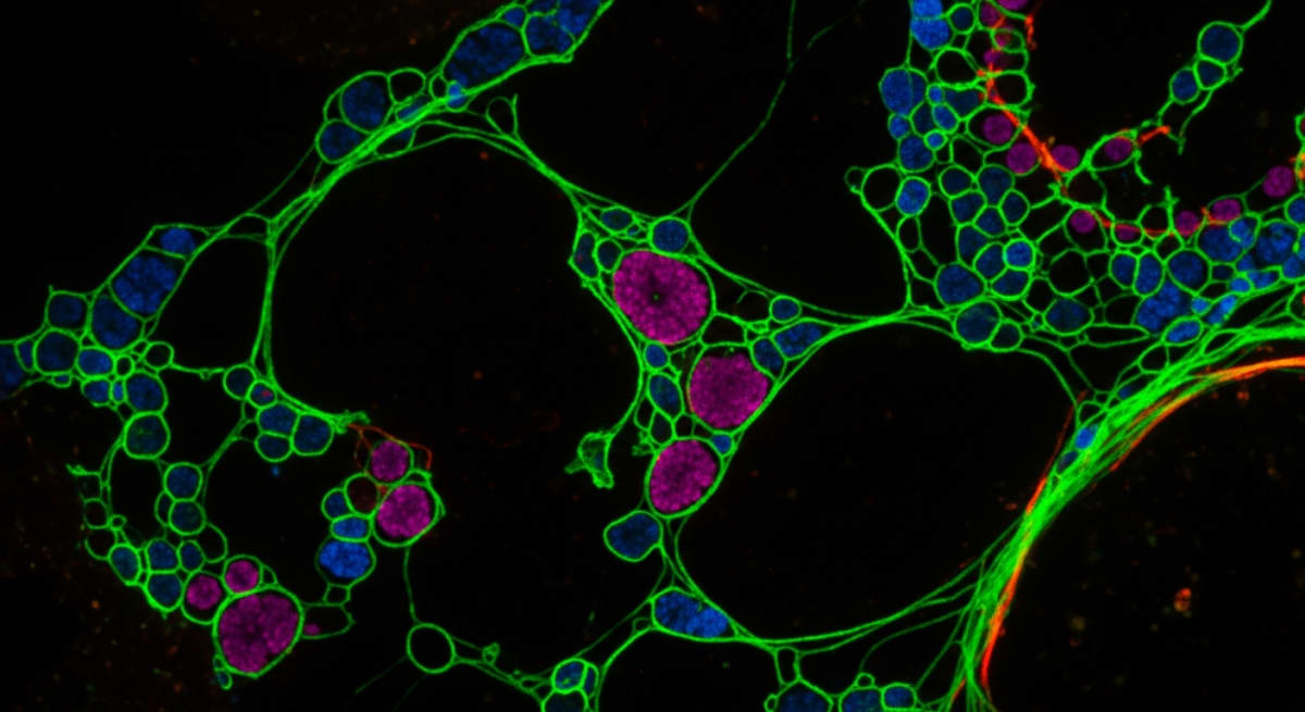
Figure: Quantitative Analysis of Peptide-Membrane Interactions Using Cell-Free Fluorescence Assays
Why Salt Removal and Cold Storage Protect Fluorescence Data
Fluorescence spectroscopy is sensitive enough to detect single-digit nanomolar shifts in light, which means the quality of the lyophilised vial directly controls the accuracy of the readout. If a peptide batch sits below 99% purity by High-Performance Liquid Chromatography (HPLC), leftover truncated chains and synthesis byproducts will float in the cuvette. These unwanted fragments carry their own electrical charges and hydrophobic patches, sticking to the synthetic liposomes and skewing the binding curves. Even worse, aromatic impurities can absorb ultraviolet light or give off background glow, ruining the signal-to-noise ratio during tryptophan or FRET measurements.
Trifluoroacetic acid (TFA) is another hidden problem in standard synthesis. Residual TFA counter-ions change the pH and ionic strength of the assay buffer and can physically disrupt delicate lipid vesicles on their own. Sourcing sequences that have undergone proper TFA salt removal—swapping harsh acid salts for gentle acetate or hydrochloride balances—keeps artificial membranes intact so the fluorescence data reflects only the target peptide. Storage habits matter just as much once the vial arrives in the lab. Heat and light slowly break down amino acid bonds and oxidise tryptophan residues, dulling their natural fluorescence. Keeping lyophilised vials sealed at 2°C to 8°C in a dark fridge, and preparing fresh aliquots with bacteriostatic reconstitution solution or ultrapure buffer right before the assay, ensures crisp, repeatable optical curves every time.
Frequently Asked Questions
Why do researchers search for cell-free fluorescence assays when studying peptide-membrane interactions?
Researchers use cell-free fluorescence assays to measure exactly how peptides bind to lipid barriers without the messy interference of whole living cells. Published studies show these artificial membrane models let scientists analyse pure binding kinetics, structural folding, and pore formation using simple light-emitting dyes and controlled protocol ratios.
What is the typical timeline for running a quantitative peptide-membrane fluorescence study?
A standard fluorescence assay takes between two and six hours from lipid preparation to final data collection. Once the artificial vesicles and dye solutions are mixed, the actual binding reaction happens in seconds, though researchers often monitor the optical signalling for thirty minutes to confirm equilibrium.
Can multiple peptides or lipids be combined in a single fluorescence assay?
Yes, scientists frequently combine two or more peptides or varied lipid types in one assay to observe competitive binding and synergistic effects. Laboratory models use distinct fluorescent tags with different colour wavelengths so researchers can track each compound independently and characterise how they alter membrane stability together.
How do peptide purity and fridge storage affect fluorescence assay accuracy?
High peptide purity above ninety-five percent and strict cold storage are essential to prevent false light signals during quantitative testing. Leftover synthesis impurities or degraded fragments can quench fluorescent dyes, so researchers store freeze-dried powders at minus twenty degrees Celsius and keep dissolved stock in the fridge briefly.
Bibliography
- Wimley WC, White SH. Experimentally determined hydrophobicity scale for proteins at membrane interfaces. Nature Structural Biology. 1996;3(10):842-848. View published research
- Ladokhin AS, Jayasinghe S, White SH. How to measure and analyze tryptophan fluorescence in membranes properly, and why bother? Analytical Biochemistry. 2000;285(2):235-245. View published research
- Parasassi T, De Stasio G, d'Ubaldo A, Gratton E. Phase fluctuation in phospholipid membranes revealed by Laurdan fluorescence. Biophysical Journal. 1990;57(6):1179-1186. View published research
- Struck DK, Hoekstra D, Pagano RE. Use of resonance energy transfer to monitor membrane fusion. Biochemistry. 1981;20(14):4093-4099. View published research
- Santos NC, Prieto M, Castanho MA. Quantifying molecular partition into model systems of biomembranes: an emphasis on optical spectroscopic methods. Biochimica et Biophysica Acta. 2003;1612(2):123-135. View published research
See our UK peptide research guides & COA briefings first in Google Search.
Add Amino Peptides UK as a Preferred Source on Google⚠️ Research Use Only Disclaimer: All peptides and compounds are sold strictly for in-vitro laboratory research purposes only. Not intended for human or veterinary use, not a dietary supplement, and not approved to diagnose, treat, cure, or prevent any disease or condition. For use by qualified researchers in suitably equipped laboratory environments only. Amino Peptides Ltd operates in full compliance with UK MHRA regulations and applicable EU research chemical guidelines.