
Structural Dynamics of GHK-Cu: Analysing Copper Chelation Equilibrium and Extracellular Matrix Gene Modulation
12th Oct 2026

Copper is a heavy metal. On its own, it is highly reactive and difficult for isolated cells to manage safely. Yet, in laboratory cell cultures, copper acts as a vital switch for cellular function. It drives the enzymes that cells use to generate energy and build structural proteins. The challenge for researchers is delivering this metal into cells without causing toxicity. Free copper ions in a petri dish can create damaging chemical reactions. This is where a specific molecular carrier comes into play.
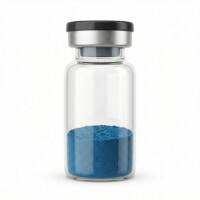
GHK-Cu
HPLC-verified lyophilised compound, UK-domestic supply. Manufactured under controlled conditions for qualified in-vitro laboratory research.
View Reagent Profile ›The compound is Glycyl-L-histidyl-L-lysine, commonly known as GHK. When it binds to a single copper ion, it forms the complex GHK-Cu. This tripeptide is a simple chain of three amino acids: glycine, histidine, and lysine. In the laboratory, researchers study how this specific molecule holds onto copper and how it interacts with isolated connective tissue cells.
Understanding how this molecule functions begins with its structure. The peptide acts like a microscopic claw. In chemistry, this gripping action is called chelation. The three amino acids fold into a specific shape that perfectly surrounds the copper ion. The histidine molecule, in particular, provides a strong chemical anchor that keeps the metal secured within the peptide chain.
This grip is not permanent. It exists in a state of delicate balance, known as equilibrium. The peptide must hold the copper tightly enough to keep it stable while floating in a liquid solution. However, it must also hold the metal loosely enough to release it when the molecule interacts with a cell membrane.
If the grip is too tight, the copper remains trapped inside the peptide and the cell cannot use it. If the grip is too loose, the copper drops out of the molecule prematurely, creating reactive oxygen species that can damage the cell culture. Laboratory analysis confirms this precise balance. Researchers verifying the purity of their compounds often review a certificate of analysis to ensure the peptide structure is completely intact before beginning any cellular assays.
Before the peptide ever reaches the petri dish, its structural integrity must be preserved through a process called lyophilisation. This is a complex freeze-drying technique that removes all water from the compound while it is frozen in a vacuum. Water is the enemy of peptide stability during storage. If moisture is present, the amino acid bonds can break down, and the copper ion can detach from the histidine anchor. The resulting lyophilised powder is highly stable, provided it remains sealed.
When researchers apply this compound to isolated fibroblasts in a controlled petri dish, they observe distinct changes in cellular behaviour. Fibroblasts are the primary cells responsible for building the extracellular matrix. This matrix is the structural framework that holds tissues together. It is made largely of tough proteins like collagen and flexible proteins like elastin.
The addition of the copper peptide alters gene expression within these isolated cells. This means the peptide signals the cell's DNA to turn certain manufacturing processes on or off. In laboratory assays, fibroblasts exposed to the compound rapidly increase their production of structural collagen.
The type of collagen produced is highly specific. In a standard cellular assay, fibroblasts do not just produce generic proteins. The copper peptide primarily signals the DNA to increase the expression of Type I and Type III collagen. Type I collagen provides rigid tensile strength, acting like microscopic steel beams within the matrix. Type III collagen provides flexibility and is often the first protein laid down during matrix remodelling. By measuring the ratio of Type I to Type III collagen in the petri dish, researchers can quantify the exact impact of the peptide on cellular manufacturing.
However, building new matrix proteins is only half of the process. The cells also increase the production of specific enzymes called matrix metalloproteinases. These enzymes act like microscopic scissors. They cut up and break down old, damaged matrix proteins floating in the cell culture.
The specific enzymes produced are known as MMP-1 and MMP-2. The peptide upregulates the expression of these enzymes in isolated cell cultures. MMP-1 specifically targets and cuts intact collagen molecules that have become misfolded or damaged. MMP-2 then breaks down these fragments further. Without this clearing process, the extracellular matrix would become cluttered with useless protein debris. The peptide ensures that the cell culture maintains a clean environment for new structural growth.
This dual action is why the molecule is heavily studied in cellular biology. It does not just stimulate new protein production. It also signals the cell to clear away cellular debris, allowing for a complete remodelling of the extracellular matrix in the petri dish. For exact molecular weights and structural data used to calibrate these controlled studies, scientists consult the product specification sheet.
When setting up these complex experiments, laboratories require highly pure reagents. Using verified materials ensures that the gene modulation observed under the microscope is actually caused by the peptide itself, and not by heavy metal contaminants or degraded amino acid chains.
It is crucial to separate laboratory observations from whole-body outcomes. A petri dish is a highly controlled, artificial environment. Isolated fibroblasts floating in a nutrient broth do not face the complex biological barriers of a living organism. They do not have a liver to filter the compound, nor an immune system to react to it.
When a peptide increases collagen expression in cultured cells, it simply means a specific chemical reaction occurred under strict laboratory conditions. You can see why the claim spread that this compound might alter human tissue. However, isolated cellular reactions do not translate directly to human biology. The data shows molecular signalling in a controlled environment, nothing more.
A cellular observation is a starting point for chemistry, not a biological conclusion.
The mechanics of how the peptide enters the cell are also a subject of intense study. The molecule does not simply smash through the cell wall. It interacts with specific receptors on the surface of the fibroblast. Once attached, the cell membrane folds inward, pulling the peptide and its copper cargo inside.
Once inside the cell, the equilibrium shifts. The internal environment of the fibroblast is different from the liquid outside. This change in chemical pressure causes the peptide to finally release its grip on the copper ion. The free copper is then immediately grabbed by internal cellular proteins.
These internal proteins act as molecular shuttles. When the tripeptide releases its copper payload just inside the cell membrane, these shuttle proteins immediately bind to the metal. They carry the copper safely through the watery interior of the cell. They deliver the metal directly to the cell's nucleus and manufacturing centres, where it is incorporated into newly synthesised enzymes and used to modulate gene expression.
This precise delivery system prevents the copper from causing damage while in transit. The tripeptide acts as a protective escort, ensuring the metal reaches the exact location needed to trigger the genetic switches for extracellular matrix production.
Frequently Asked Questions in Laboratory Research
What is the standard concentration for a ghk cu 1 solution in cellular assays?
In many baseline in-vitro studies, researchers prepare a 1 percent concentration of the peptide. A 'ghk cu 1' solution provides a standard molarity for observing gene modulation in isolated fibroblasts. This specific concentration allows scientists to measure changes in collagen production without overwhelming the cell cultures with excess copper, which could lead to cellular toxicity. A 1 percent baseline provides a reproducible standard, allowing different laboratories to compare their results accurately.
Why do researchers avoid ghk cu copper peptide tablets for in-vitro studies?
Cellular assays require precise, direct application of a pure compound to isolated cells. 'Ghk cu copper peptide tablets' contain binders, fillers, and digestive coatings designed for oral breakdown. These commercial additives would instantly contaminate a petri dish. They would ruin the controlled environment required for accurately measuring extracellular matrix changes under a microscope. Laboratory research demands absolute precision, and tablet formats introduce unknown variables into the assay.
How do laboratories source ghk cu copper peptide uk materials?
When searching for 'ghk cu copper peptide uk', researchers must look for chemical suppliers that provide strict analytical testing. The copper chelation bond is fragile and easily disrupted by poor manufacturing. Sourcing verified materials ensures the molecule has not degraded during transit or storage, guaranteeing that the structural dynamics remain intact for the experiment. High-quality suppliers provide detailed mass spectrometry results to prove the exact molecular weight of the compound.
Is there a difference when searching for ghk cu peptide uk versus the copper-bound version?
Yes, there is a significant chemical difference. The base GHK molecule can exist without the copper ion attached. When laboratories look for 'ghk cu peptide uk', they are specifically seeking the chelated form. The raw, unchelated GHK molecule behaves differently in cellular assays and does not trigger the same extracellular matrix gene modulation as the copper-bound complex. The unchelated peptide can act as a scavenger, unintentionally altering the baseline chemistry of the petri dish.
What are the storage requirements for ghk cu uk research supplies?
For any 'ghk cu uk' laboratory inventory, the lyophilised powder must be stored in a freezer, strictly away from light and moisture. Once the powder is reconstituted with a bacteriostatic reconstitution solution, the resulting liquid must be kept refrigerated. Proper temperature control is the only way to maintain the structural dynamics and chelation equilibrium of the peptide over time. If left at room temperature, the amino acid chain will break apart, and the copper will precipitate out of the solution.
The structural dynamics of this tripeptide offer a clear window into the mechanics of cellular signalling. By acting as a precise molecular carrier, the compound successfully delivers copper to isolated fibroblasts, triggering a complex cascade of genetic changes. The equilibrium of its molecular grip dictates its entire effectiveness in the laboratory setting.
Research into extracellular matrix gene modulation continues to map out exactly how these chemical switches operate at the molecular level. The data remains strictly confined to the petri dish, providing a foundational understanding of peptide chemistry. As laboratories continue to analyse the chelation equilibrium, the exact mechanisms of copper delivery and cellular response become increasingly clear.

- Pickart, L., & Margolina, A. (2018). Regenerative and Protective Actions of the GHK-Cu Peptide in the Light of the New Gene Data. International Journal of Molecular Sciences, 19(7), 1987. View published research
- Pickart, L. (2008). The human tri-peptide GHK and tissue remodeling. Journal of Biomaterials Science, Polymer Edition, 19(8), 969-988. View published research
- Maquart, F. X., Pickart, L., Laurent, M., Gillery, P., Monboisse, J. C., & Borel, J. P. (1988). Stimulation of collagen synthesis in fibroblast cultures by the tripeptide-copper complex glycyl-L-histidyl-L-lysine-Cu2+. FEBS Letters, 238(2), 343-346. View published research
- Siméon, A., Wegrowski, Y., Bonté, F., & Maquart, F. X. (2000). Expression of glycosaminoglycans and small proteoglycans in wounds: modulation by the tripeptide-copper complex glycyl-L-histidyl-L-lysine-Cu(2+). Journal of Investigative Dermatology, 115(6), 962-968. View published research
- Kang, Y. A., Choi, H. R., Na, J. I., Huh, C. H., Kim, H. R., Park, K. C., & Youn, S. W. (2009). Copper-GHK increases integrin expression and p63 positivity by keratinocytes. Archives of Dermatological Research, 301(4), 301-306. View published research
- Gruchlik, A., Jurzak, M., Chodurek, E., & Dzierzewicz, Z. (2012). Effect of GHK-Cu on proliferation of normal human dermal fibroblasts in vitro. Acta Poloniae Pharmaceutica, 69(6), 1303-1306. View published research
⚠️ Research Use Only Disclaimer: All peptides and compounds are sold strictly for in-vitro laboratory research purposes only. Not intended for human or veterinary use, not a dietary supplement, and not approved to diagnose, treat, cure, or prevent any disease or condition. For use by qualified researchers in suitably equipped laboratory environments only. Amino Peptides Ltd operates in full compliance with UK MHRA regulations and applicable EU research chemical guidelines.
Independent, batch-specific documentation for GHK-Cu — reviewed prior to publication for in-vitro laboratory research and molecular stability reference.