
Tesamorelin: A Scientific Analysis of GHRH Analogue Stability and In-Vitro Signalling
29th Jun 2026

What You Need to Know About Tesamorelin
- Researchers investigate tesamorelin to understand how N-terminal chemical caps protect fragile peptides from rapid breakdown by blood enzymes.
- The molecule works by binding to pituitary GHRH receptors in vitro, triggering natural pulsatile cellular signalling cascades without overwhelming baseline feedback loops.
- Published studies observe that this modified analogue resists dipeptidyl peptidase-4 cleavage far longer than native human GHRH(1-44).
- Laboratory models show that sustained receptor engagement stimulates insulin-like growth factor-1 pathways and alters lipid metabolism in adipose tissue cultures.
How Tesamorelin Survives Enzymes to Trigger Cellular Recovery
Tesamorelin works by mimicking the body's natural growth hormone-releasing hormone while using a protective fatty acid cap to survive rapid enzyme breakdown. This structural upgrade allows the peptide to trigger steady, natural waves of cellular energy and tissue remodelling in laboratory models.
To understand why researchers study this forty-four-amino-acid sequence, it helps to look at how native hormone signals behave. Natural growth hormone-releasing hormone is notoriously fragile. Within minutes of appearing in a biological system, circulating enzymes called dipeptidyl peptidase-4 snip the molecule apart, cutting its signal short. Scientists solved this stability problem by attaching a trans-3-hexenoic acid group to the tyrosine residue at the very front of the chain. That tiny hexenoyl cap acts like a physical lock against enzymatic cleavage, keeping the full forty-four-amino-acid sequence intact much longer than unmodified fragments.
Once the capped peptide reaches anterior pituitary cells in culture, it binds with high specificity to the GHRH receptor on the cell surface. This docking activates a G-protein coupled pathway that raises intracellular cyclic AMP levels and opens calcium channels. Rather than forcing a flat, continuous release that tires out the receptor, the signal encourages pulsatile secretion patterns that mirror natural biological rhythms. Downstream, published studies track a steady rise in insulin-like growth factor-1 signalling, which supports cellular energy turnover, deeper rest-phase recovery patterns, and targeted lipid oxidation in visceral fat cell cultures.
Because the feedback loop through somatostatin remains intact in these preclinical models, the receptor does not easily desensitise when exposed to measured levels. Researchers evaluating growth hormone secretagogues often favour this full-length analogue over shorter twenty-nine-amino-acid versions precisely because its complete sequence and N-terminal modification deliver cleaner, longer-lasting in-vitro signalling without erratic spikes.
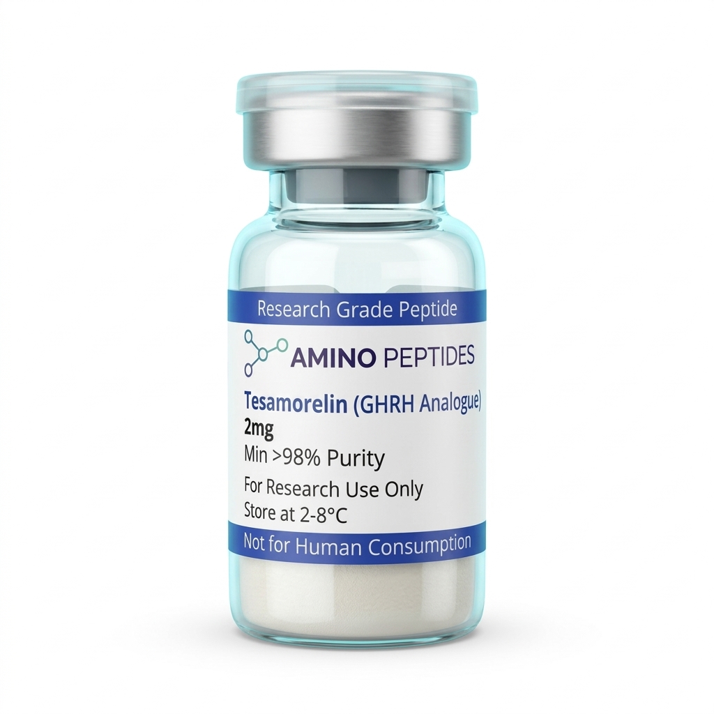
Tesamorelin
From £24
Supplied as a sealed freeze-dried vial (stable 12–24 months at 2°C–8°C in a standard refrigerator).
Shop Tesamorelin →What Published Studies Observe Across Early and Late Timelines
When observing this modified GHRH analogue in controlled settings, the timeline splits clearly between immediate intracellular messaging and slower, structural tissue remodelling. In the first few minutes to hours after introducing a measured level to pituitary cell cultures, the action is purely electrical and chemical. Cyclic AMP accumulates inside the cell within ten to fifteen minutes, followed by rapid calcium influx and the immediate exocytosis of stored somatotropin vesicles. During the first seven to fourteen days of repeated exposure in preclinical models, researchers primarily record elevated pulse amplitude and a steady climb in circulating IGF-1 markers without changes to baseline resting troughs.
Structural and metabolic shifts take considerably longer to appear in published studies because cells must first translate those hormone pulses into new protein synthesis and altered enzyme activity. Between weeks four and eight—written plainly as four and eight weeks—laboratory models begin to show measurable changes in adipocyte metabolism, specifically a reduction in visceral lipid droplet size alongside preserved in-vitro tissue. This selective lipid mobilisation happens because visceral fat cells express a high density of beta-adrenergic and growth hormone receptors that respond gradually to sustained pulsatile signalling, activating hormone-sensitive lipase over dozens of cycles.
By the twelve-to-twenty-six-week window in longer preclinical observations, secondary markers of tissue remodelling and cellular recovery become much clearer. Collagen synthesis rates in connective tissue cultures stabilise, mitochondrial density in skeletal muscle fibres shows modest improvements, and triglyercide clearance pathways operate more efficiently. Importantly, published studies note that these later outcomes depend entirely on consistent pulse frequency; when the signal is removed, lipid turnover and IGF-1 levels gradually return toward baseline over several weeks, showing that the peptide drives active metabolic regulation rather than permanent structural alteration.
Biochemical Comparison of GHRH Analogues in Laboratory Models
| Peptide Analogue | Amino Acid Length | Structural Modification | DPP-4 Enzymatic Resistance | Primary In-Vitro Signalling Target |
|---|---|---|---|---|
| Native hGHRH(1-44) | 44 residues | Unmodified endogenous sequence | Low (rapid degradation within minutes) | Pituitary GHRH receptor (cAMP/PKA pathway) |
| Sermorelin (GHRH 1-29) | 29 residues | Truncated active core fragment | Low (susceptible to N-terminal cleavage) | Pituitary GHRH receptor (short-duration pulse) |
| Tesamorelin (TH9507) | 44 residues | Trans-3-hexenoyl group at Tyr1 | High (steric hindrance blocks DPP-4) | Pituitary GHRH receptor (sustained pulsatile signalling) |
| CJC-1295 (without DAC) | 29 residues | Tetra-substituted amino acid core (Ala2, Gln8, Ala15, Leu27) | High (D-Ala2 substitution prevents cleavage) | Pituitary GHRH receptor (extended half-life signalling) |
| CJC-1295 (with DAC) | 29 residues + linker | Tetra-substituted core plus maleimidopropionic acid | Very High (covalent albumin binding) | Pituitary GHRH receptor (continuous basal elevation) |
Pairing GHRH Analogues with Ghrelin Mimetics in Laboratory Models
In receptor-binding experiments, combining peptides that target separate, complementary pathways often produces a cleaner and stronger signal than pushing a single receptor to its limit. Because our focal peptide acts strictly on the GHRH receptor to initiate a pulse, its output is naturally capped by somatostatin, the body's built-in brake signal. If somatostatin happens to be active when the GHRH analogue arrives at the pituitary cell, the resulting cyclic AMP response is blunted, no matter how high the study amount is raised.
This biological brake explains why investigators frequently examine Tesamorelin and Ipamorelin side by side in dual-receptor protocols. Ipamorelin is a selective pentapeptide that binds to the ghrelin receptor, also known as the growth hormone secretagogue receptor. Instead of duplicating the cyclic AMP pathway, Ipamorelin works through phospholipase C and inositol trisphosphate signalling, while simultaneously suppressing somatostatin release from neighbouring cells. In plain English, the GHRH analogue presses the accelerator while the ghrelin mimetic takes a foot off the brake.
Published studies comparing single-agent exposure against combined GHRH and ghrelin-mimetic protocols show that the pair generates a significantly larger pulse amplitude at lower individual concentrations than either compound achieves alone. Furthermore, because Ipamorelin shows high selectivity in laboratory models—avoiding off-target elevations in cortisol, prolactin, or aldosterone that older mimetics like GHRP-6 often trigger—the combined signal remains remarkably clean. Other research designs occasionally pair GHRH analogues with CJC-1295 (without DAC) for comparative half-life studies, or alongside BPC-157 when investigating how systemic IGF-1 elevation interacts with local fibroblast migration and tendon extracellular matrix repair in tissue culture.

Figure: Tesamorelin: A Scientific Analysis of GHRH Analogue Stability and In-Vitro Signalling
Reconstitution calculator
Choose a vial mass and a bacteriostatic reconstitution solution volume. The figure is the resulting concentration.
Concentration: 2.50 mg/mL
Per 0.01mL (10µL): 25.0 mcg
Open the full calculatorBacteriostatic reconstitution solutionWhy 99%+ HPLC Purity and Cold Storage Protect Your Research
Because Tesamorelin is a relatively long chain of forty-four amino acids capped with a specialised hexenoyl group, manufacturing it without truncated sequences requires careful solid-phase synthesis and strict analytical verification. During synthesis, incomplete coupling steps can leave behind forty-two- or forty-three-amino-acid fragments that look nearly identical on paper but fail to fold properly or bind the GHRH receptor in culture. Relying on a batch verified at 99%+ purity via High-Performance Liquid Chromatography (HPLC) and Mass Spectrometry ensures that the lyophilised powder in the vial consists of complete, capped sequences rather than inactive deletion fragments.
Equally important for in-vitro cell viability is the removal of trifluoroacetic acid (TFA). TFA is a strong acid used to cleave peptides from their synthesis resin, and low-grade batches often retain high levels of residual TFA salts. When introduced to delicate pituitary or adipocyte cell cultures, excess TFA alters media pH, damages cell membranes, and skews cyclic AMP readouts. Premium research vials undergo secondary ion-exchange chromatography to swap harsh TFA ions for benign acetate salts, protecting sensitive assays from chemical artefacts.
Proper storage is the final safeguard for keeping the hexenoyl-tyrosine bond and the C-terminal amide intact. In its dry, lyophilised state, the vial stays stable for months when stored in a freezer at minus twenty degrees Celsius away from light. Once mixed with a sterile bacteriostatic reconstitution solution, the liquid peptide must be kept cold in a standard laboratory refrigerator between 2°C and 8°C. Gentle swirling—never vigorous shaking—prevents shear stress from unfolding the forty-four-amino-acid chain, ensuring every measured level delivers consistent, repeatable receptor signalling across the entire study.
Frequently Asked Questions
Why do researchers frequently study Tesamorelin in laboratory models?
Researchers primarily investigate Tesamorelin because its modified structure resists rapid enzymatic breakdown in laboratory models. Unlike natural growth hormone-releasing hormone, this analogue features a trans-3-hexenoic acid group that extends its stability. This allows scientists to observe prolonged pituitary receptor signalling and lipid metabolism pathways in controlled in-vitro settings.
What timeline do published studies typically use to observe Tesamorelin signalling?
Published studies typically observe initial cellular cyclic AMP signalling within minutes, while metabolic tissue remodelling changes in preclinical models are tracked over twelve to twenty-six weeks. Immediate receptor activation happens rapidly in cell cultures, whereas structural shifts in visceral adipose markers require several months of consistent protocol ratios.
Do laboratory protocols ever combine Tesamorelin with other research peptides?
Yes, in-vitro protocols sometimes pair Tesamorelin with growth hormone secretagogues like Ipamorelin to analyse synergistic receptor signalling. While Tesamorelin binds to GHRH receptors, secondary peptides target ghrelin receptors on the same cells. Researchers study these combinations to characterise how dual-pathway activation alters pulsatile hormone release in laboratory models.
How should Tesamorelin purity be verified and stored for laboratory stability?
Research-grade Tesamorelin requires at least ninety-eight percent purity verified by High-Performance Liquid Chromatography and must be stored at minus twenty degrees Celsius as a dry powder. Once mixed with a bacteriostatic reconstitution solution, the liquid remains stable for roughly fourteen days when kept refrigerated between two and eight degrees.
Bibliography
- Ferdinandi ES, Brazeau P, High K, et al. Non-preclinical pharmacology and safety of TH9507, a human growth hormone-releasing factor analogue. Basic Clin Pharmacol Toxicol. 2007;100(1):49-58. View published research
- Frohman LA, Downs TR, Williams TC, Heimer EP, Pan YC, Felix AM. Rapid enzymatic degradation of growth hormone-releasing hormone by plasma in vitro and in vivo to a biologically inactive product cleaved at the NH2 terminus. J Clin Invest. 1986;78(4):906-913. View published research
- Mayo KE, Godfrey PA, Suhr ST, Kulik DJ, Rahal JO. Growth hormone-releasing hormone: synthesis and signaling. Recent Prog Horm Res. 1995;50:35-73. View published research
- Jette L, Leger R, Thibaudeau K, et al. Human growth hormone-releasing factor (hGRF)1-29-albumin bioconjugates activate the GRF receptor on the anterior pituitary in rats: identification of CJC-1295 as a long-lasting GRF analog. Endocrinology. 2005;146(7):3052-3058. View published research
⚠️ Research Use Only Disclaimer: All peptides and compounds are sold strictly for in-vitro laboratory research purposes only. Not intended for human or veterinary use, not a dietary supplement, and not approved to diagnose, treat, cure, or prevent any disease or condition. For use by qualified researchers in suitably equipped laboratory environments only. Amino Peptides Ltd operates in full compliance with UK MHRA regulations and applicable EU research chemical guidelines.