
The Molecular Science of Tesamorelin: An In-Vitro Analysis of GHRH Receptor Activation
2nd Jul 2026
What You Need to Know About Tesamorelin
- Researchers search for this compound to understand how adding a simple hexenoyl group protects the 44-amino-acid chain from rapid enzymatic breakdown by dipeptidyl peptidase-4 (DPP-4).
- In plain English, the molecule acts like a master key that fits into GHRH receptors on pituitary cells, flipping an internal switch that tells the cell to release stored growth hormone in natural pulses.
- Published studies in laboratory models show that this receptor activation boosts cyclic adenosine monophosphate (cAMP) levels and stimulates insulin-like growth factor-1 (IGF-1) pathways without disrupting standard somatostatin feedback loops.
- Biochemists also analyse this analogue alongside truncated fragments like Sermorelin and CJC-1295 to compare how full-length sequences versus modified short chains influence receptor binding affinity and stability.
How a Protected 44-Amino-Acid Sequence Triggers Somatotroph Signalling
Tesamorelin is a synthetic 44-amino-acid peptide modified with a trans-3-hexenoic acid group that binds directly to growth hormone-releasing hormone receptors in laboratory models. This structural cap protects the sequence from rapid enzymatic breakdown while triggering natural, pulsatile cellular signalling for tissue recovery and lipid metabolism.
Native growth hormone-releasing hormone is notoriously fragile in a dish. The moment it meets the enzyme dipeptidyl peptidase-4, known simply as DPP-4, the molecule is clipped at its N-terminus and loses its biological spark within minutes. Chemists solved this problem by attaching a six-carbon fatty acid chain—trans-3-hexenoic acid—onto the tyrosine residue at position one. That small hydrophobic tail acts like a molecular bumper. It blocks DPP-4 from latching onto the cleavage site while keeping the full 44-amino-acid backbone intact so it can dock cleanly with the GHRH receptor.
Once Tesamorelin settles into the extracellular pocket of the GHRH receptor on cultured anterior pituitary somatotrophs, the seven-transmembrane protein changes shape. Inside the cell, a Gs alpha subunit splits off and switches on adenylyl cyclase. That enzyme rapidly converts adenosine triphosphate into cyclic adenosine monophosphate, or cAMP. Rising cAMP levels wake up protein kinase A, which then opens voltage-gated calcium channels along the cell membrane. Calcium ions rush inward, prompting secretory vesicles to fuse with the outer membrane and release stored somatotropin in measured waves.
Published studies favour this full-length 44-amino-acid architecture over shorter 29-amino-acid fragments such as Sermorelin because the C-terminal tail adds extra hydrogen-bonding contacts. Those extra anchor points tighten receptor binding in vitro and sustain intracellular cAMP accumulation for longer intervals. Instead of forcing a flat, continuous output, the receptor stays responsive to the negative feedback of somatostatin. In preclinical models examining cellular energy and visceral fat metabolism, this rhythmic pattern encourages lipolysis and mitochondrial fatty acid oxidation without exhausting the pituitary cell surface.
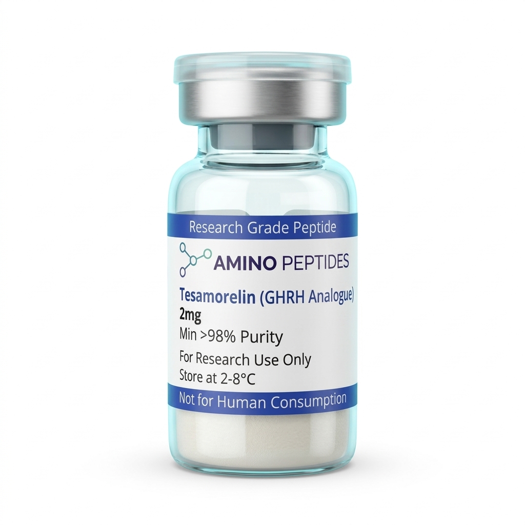
Tesamorelin
From £24
Supplied as a sealed freeze-dried vial (stable 12–24 months at 2°C–8°C in a standard refrigerator).
Shop Tesamorelin →From Rapid cAMP Spikes to Long-Term Lipid Remodelling in Preclinical Models
Laboratory timelines for Tesamorelin split neatly into two phases: the immediate electrical and chemical spark inside pituitary cultures, and the slower metabolic and structural shifts observed across multi-week preclinical models. Because the peptide works by nudging an upstream receptor rather than supplying exogenous hormone directly, the clock runs at the speed of natural gene transcription and protein synthesis.
During the first one to five minutes of in-vitro exposure, GHRH receptor occupancy hits its stride. Adenylyl cyclase activity climbs sharply, and intracellular cAMP concentrations peak within fifteen to thirty minutes at nanomolar study amounts. Calcium imaging in cultured somatotrophs shows rhythmic oscillations starting almost immediately, followed by measurable somatotropin release into the surrounding medium over the first sixty to ninety minutes. Even with the protective trans-3-hexenoic acid cap, free Tesamorelin is gradually cleared over several hours, yet the downstream messenger wave it sets in motion keeps rolling.
Between hours six and twenty-four, the second phase begins as released somatotropin engages hepatic and peripheral receptors in co-culture or animal models. This secondary handshake switches on the JAK2-STAT5 signalling pathway, driving the transcription of insulin-like growth factor-1 (IGF-1). Steady rises in secreted IGF-1 and IGF-binding protein-3 typically stabilise between days three and seven of repeated daily exposures in preclinical models.
Structural tissue remodelling takes considerably longer to show up under the microscope. In adipose cell cultures and rodent metabolic studies, shifts in hormone-sensitive lipase activity and reduced de novo lipogenesis become measurable after two to four weeks of consistent protocol ratios. Significant reductions in visceral adipocyte volume, alongside improved triglyceride clearance and collagen matrix reorganisation, generally require six to twelve weeks of sustained observation. Researchers tracking sleep-related slow-wave electrical patterns or peripheral nerve regeneration in laboratory models likewise note that transcription-driven structural changes follow this multi-week arc rather than overnight shifts.
Structural and Biochemical Comparison of GHRH Analogues in Laboratory Models
| Biochemical Feature | Tesamorelin (TH9507) | Native Human GHRH (1-44) | Sermorelin (GHRH 1-29) | Modified GRF (1-29) / CJC-1295 |
|---|---|---|---|---|
| Amino Acid Length | 44 amino acids (full-length sequence) | 44 amino acids (endogenous full-length) | 29 amino acids (N-terminal active fragment) | 29 amino acids (tetrasubstituted fragment) |
| Structural Modification | Trans-3-hexenoyl group attached to Tyr-1 at the N-terminus | Unmodified native polypeptide chain with amidated C-terminus | Truncated chain retaining the core receptor-binding domain | Four amino acid substitutions (positions 2, 8, 15, 27) with optional DAC linker |
| DPP-4 Enzymatic Resistance | High resistance due to N-terminal hexenoyl steric shielding | Very low; rapidly cleaved at the Ala-2 position in vitro | Low; susceptible to rapid enzymatic degradation in serum | High resistance due to D-Ala-2 substitution blocking peptidase cleavage |
| Primary Receptor Target | Pituitary GHRH receptor (Class B GPCR) | Pituitary GHRH receptor (Class B GPCR) | Pituitary GHRH receptor (Class B GPCR) | Pituitary GHRH receptor (Class B GPCR) |
| Dominant Second Messenger | Adenylate cyclase activation leading to cAMP and PKA signalling | Adenylate cyclase activation leading to cAMP and PKA signalling | Adenylate cyclase activation leading to cAMP and PKA signalling | Adenylate cyclase activation leading to cAMP and PKA signalling |
Pairing GHRH Analogues with Ghrelin Mimetics and Tissue Peptides in the Lab
In receptor pharmacology, pairing two molecules that hit separate but cooperative pathways often reveals how cells coordinate complex metabolic work. Tesamorelin occupies the GHRH receptor, which raises cAMP through the Gs protein pathway. However, pituitary somatotrophs also carry a completely separate docking station called the growth hormone secretagogue receptor (GHS-R1a), which responds to ghrelin and synthetic mimetics like Ipamorelin.
When researchers combine Tesamorelin with a selective ghrelin mimetic such as Ipamorelin in vitro, two distinct intracellular messengers fire at once. While Tesamorelin drives the cAMP and protein kinase A route, Ipamorelin activates phospholipase C and inositol trisphosphate, releasing calcium from internal endoplasmic reticulum stores and simultaneously suppressing somatostatin tone. Removing the somatostatin brake while pressing the GHRH accelerator produces a pulse of somatotropin release that exceeds what either peptide achieves alone at triple the concentration. Because Ipamorelin is selective and avoids spilling over into cortisol or prolactin pathways in laboratory models, the combined readout stays clean and easy to interpret.
Beyond pituitary co-stimulation, investigators frequently examine how upstream somatotropic signals interact with local extracellular matrix peptides during tissue recovery studies. While Tesamorelin-driven IGF-1 transcription supports systemic amino acid uptake and fibroblast proliferation, researchers studying dermal architecture often buy ghk-cu to observe localised copper-dependent collagen synthesis and metalloproteinase balance in parallel fibroblast assays. Similarly, surface-focused cosmeceutical assays may pair matrix-remodelling work with the snap 8 peptide to compare deep structural protein synthesis against SNARE-complex modulation at neuromuscular junctions.
Another common laboratory pairing teams Tesamorelin with CJC-1295 (without DAC) or pentadecapeptide BPC-157 in tendon and muscle regeneration models. In these multi-agent setups, the GHRH analogue supplies the steady anabolic transcription signal while the local peptide supports angiogenesis and actin filament organisation at the site of cellular stress.
Reconstitution calculator
Choose a vial mass and a bacteriostatic reconstitution solution volume. The figure is the resulting concentration.
Concentration: 2.50 mg/mL
Per 0.01mL (10µL): 25.0 mcg
Open the full calculatorBacteriostatic reconstitution solutionWhy 99%+ Purity, TFA Removal, and Cold Storage Protect Experimental Data
A 44-amino-acid chain capped with a trans-3-hexenoic acid group is a long, demanding sequence to assemble on a solid-phase resin. Every single coupling step must approach complete efficiency, or truncated deletion sequences—missing a single tyrosine, alanine, or arginine—will accumulate in the batch. If those nearly identical 42- or 43-amino-acid fragments remain in the vial, they can still bind the GHRH receptor pocket without flipping the Gs alpha switch, acting as silent competitive blockers that ruins the cAMP readout.
That is why verifying a minimum of 99% purity via High-Performance Liquid Chromatography (HPLC) and confirming the exact molecular mass of 5135.9 Daltons by Mass Spectrometry (MS) is essential before starting any assay. High-grade batches also undergo ion-exchange resin polishing to strip out trifluoroacetic acid (TFA). Leftover TFA salts from cleavage are acidic and cytotoxic to delicate pituitary primary cultures; swapping TFA for a benign acetate counter-ion keeps the pH of the culture medium stable and prevents premature membrane damage.
Once a verified lyophilised cake arrives in the lab, temperature control protects that chemical architecture. Unreconstituted vials should be stored at -20°C away from bright light, where the dry peptide lattice stays stable for months. When preparing a working stock with a sterile bacteriostatic reconstitution solution, the liquid should be trickled gently down the inside glass wall rather than sprayed straight onto the powder. Vigorous shaking creates air-water interfaces that can shear or unfold the 44-amino-acid spiral; slow, gentle swirling dissolves the cake cleanly.
After reconstitution, the vial belongs in a dedicated laboratory fridge maintained strictly between 2°C and 8°C. At these cool temperatures, hydrolysis and oxidation of sensitive methionine and tyrosine residues are kept to a minimum for up to three to four weeks. Avoiding repeated freeze-thaw cycles by aliquoting the solution into smaller single-run volumes ensures that the last assay of the month performs identically to the first.
Frequently Asked Questions
Why do researchers study Tesamorelin in laboratory models?
Researchers study Tesamorelin because its modified molecular structure resists rapid enzymatic breakdown while activating growth hormone-releasing hormone receptors in vitro. Published studies analyse how this synthetic forty-four amino acid chain triggers specific intracellular signalling pathways. Scientists use it to characterise receptor binding affinity and cellular responses in controlled laboratory models.
What timeline do published studies observe for receptor signalling?
In vitro assays show that receptor binding and initial cyclic AMP signalling occur within minutes of introducing the peptide. Longer preclinical models evaluating downstream cellular recovery or tissue remodelling markers typically run for several weeks. The exact observation window depends entirely on the specific biological pathway researchers choose to analyse.
Can Tesamorelin be combined with other peptides in research protocols?
Yes, laboratory protocols frequently pair Tesamorelin with ghrelin mimetics like Ipamorelin to investigate synergistic receptor signalling. While Tesamorelin targets the GHRH receptor, secondary compounds bind to separate receptors on the same cells. Published studies examine these combined protocol ratios to characterise amplified signalling outputs in isolated pituitary cell cultures.
How should lyophilised Tesamorelin be stored to maintain high purity?
Lyophilised Tesamorelin must be stored in a freezer at minus twenty degrees Celsius to preserve its structural purity. Once mixed with a sterile bacteriostatic reconstitution solution for an assay, the liquid sample should be kept refrigerated between two and eight degrees Celsius and utilised promptly to prevent molecular degradation.
Bibliography
- Ferdinandi ES, Brazeau P, High K, Procter B, Fennell S, Dubreuil P. Non-preclinical pharmacology and safety evaluation of TH9507, a human growth hormone-releasing factor analogue. Basic Clin Pharmacol Toxicol. 2007;100(1):49-58. View published research
- Mayo KE, Godfrey PA, Suhr ST, Kulik DJ, Rahal JO. Growth hormone-releasing hormone: synthesis and signaling. Recent Prog Horm Res. 1995;50:35-73. View published research
- Frohman LA, Downs TR, Williams TC, Heimer EP, Pan YC, Felix AM. Rapid enzymatic degradation of growth hormone-releasing hormone by plasma in vitro and in vivo to a biologically inactive product cleaved at the NH2 terminus. J Clin Invest. 1986;78(4):906-913. View published research
- Zhou F, Zhang H, Cong Z, Zhao LH, Zhou Q, Mao C, Cheng X, Shen DD, Cai X, Ma C, Wang Y, Jiang Y, Jiang H, Zhang Y, Xu HE, Wang MW. Structural basis for activation of the growth hormone-releasing hormone receptor. Nat Commun. 2020;11(1):5205. View published research
⚠️ Research Use Only Disclaimer: All peptides and compounds are sold strictly for in-vitro laboratory research purposes only. Not intended for human or veterinary use, not a dietary supplement, and not approved to diagnose, treat, cure, or prevent any disease or condition. For use by qualified researchers in suitably equipped laboratory environments only. Amino Peptides Ltd operates in full compliance with UK MHRA regulations and applicable EU research chemical guidelines.