
Understanding Tesamorelin Receptors: In-Vitro Signaling and Receptor Kinetics
24th Jul 2026

What You Need to Know About Tesamorelin
- Researchers search for this compound to understand how adding a six-carbon fatty acid chain protects the native 44-amino-acid hormone sequence from rapid enzymatic breakdown in serum.
- In simple terms, the molecule acts like a precision key that locks onto pituitary surface receptors, flipping an internal switch that tells the cell to release stored growth hormone in natural waves.
- Published laboratory studies observed that this modified structure binds to target receptors with high affinity while resisting cleavage by dipeptidyl peptidase-4 (DPP-4) enzymes far longer than unmodified peptides.
- Preclinical models show that activating this specific receptor triggers cyclic AMP accumulation and calcium influx without disrupting baseline feedback loops controlled by somatostatin.
How Tesamorelin Locks Onto Pituitary Receptors to Drive Cellular Recovery
Tesamorelin binds directly to growth hormone-releasing hormone receptors on anterior pituitary cells, triggering natural, rhythmic pulses of growth hormone release that support cellular energy and tissue recovery. A small fatty acid cap attached to the N-terminus shields the chain from rapid enzyme breakdown, giving it a much longer active window at the receptor site than native GHRH.
When exploring Understanding Tesamorelin Receptors: In-Vitro Signaling and Receptor Kinetics, the story starts with a forty-four-amino-acid sequence that mirrors our body's own growth hormone-releasing hormone. Native GHRH breaks down in the bloodstream in just a few minutes because an enzyme called dipeptidyl peptidase-4 snips the chain right between the second and third amino acids. Chemists solved this rapid breakdown by attaching a trans-3-hexenoyl group to the tyrosine residue at position one. That hydrophobic tail acts like a molecular bumper, blocking the enzyme while helping the peptide dock tightly into the GHRH receptor pocket.
Once Tesamorelin clicks into that seven-transmembrane G-protein-coupled receptor on somatotroph cells, it flips an internal switch. The alpha-s subunit separates and switches on adenylyl cyclase, which converts adenosine triphosphate into cyclic adenosine monophosphate, or cAMP. Rising cAMP levels wake up Protein Kinase A. This enzyme opens voltage-gated calcium channels along the cell membrane, letting calcium ions rush inside. That sudden calcium spark causes storage vesicles packed with growth hormone to fuse with the outer membrane and release their contents. Because this pathway relies on the cell's natural feedback loops, somatostatin can still step in to apply the brakes. Published studies show that this preserves the normal wave-like rhythm of hormone output rather than forcing a flat, constant flood, which explains why laboratory models connect this cleaner kinetic profile with steady nocturnal recovery, deeper slow-wave sleep patterns, and visceral lipid mobilisation.
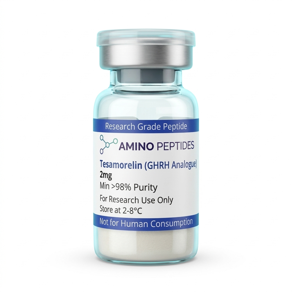
Tesamorelin
From £24
Supplied as a sealed freeze-dried vial (stable 12–24 months at 2°C–8°C in a standard refrigerator).
Shop Tesamorelin →From First-Minute Receptor Kinetics to Twelve-Week Structural Shifts
In laboratory models, the gap between the very first receptor contact and measurable structural remodelling unfolds across three distinct stages. Watching how fast the molecule binds, signals, and clears helps researchers separate immediate second-messenger spikes from slower changes in lipid metabolism and tissue recovery.
Stage One: Immediate In-Vitro Signalling (Zero to Sixty Minutes). Within seconds of introducing a measured level of Tesamorelin to cultured pituitary somatotrophs, receptor kinetics kick into high gear. Dissociation constant measurements show that the hexenoyl-modified peptide locks onto the GHRH receptor with high selectivity, showing virtually zero spillover onto related secretin-family receptors. Intracellular cAMP concentrations climb sharply inside the first five to ten minutes. Calcium influx and the first wave of growth hormone exocytosis peak between fifteen and forty-five minutes, followed by natural receptor internalisation and recycling so the cell can reset for the next pulse.
Stage Two: Downstream Hepatic and Metabolic Shifts (Weeks One to Four). Over the first several weeks of repeated pulsatile activation in preclinical models, those regular growth hormone waves travel to the liver and stimulate insulin-like growth factor-1 production. Unlike flat, continuous receptor stimulation—which quickly desensitises the pituitary—pulsatile kinetics keep receptor density stable. By week two, circulating IGF-1 levels reach a steady plateau. By week four, heightened hormone-sensitive lipase activity starts breaking down stored triglycerides inside visceral fat depots without disturbing in-vitro fat pads or impairing glucose balance.
Stage Three: Macroscopic Tissue Remodelling (Weeks Eight to Twenty-Six). Long-term published studies tracking receptor-driven outcomes over two to six months record clear structural shifts. Deep visceral adipose tissue volume drops measurably between weeks eight and twelve as mitochondrial fatty acid oxidation stays elevated. Over twelve to twenty-six weeks, models also show improved collagen turnover, denser dermal structure, and enhanced peripheral nerve regeneration markers driven by sustained, physiological IGF-1 signalling.
Biochemical Comparison of GHRH Analogues and Secretagogue Receptor Ligands in Laboratory Models
| Compound | Amino Acid Length | Primary Receptor Target | Key Structural Modification | Primary Intracellular Messenger |
|---|---|---|---|---|
| Tesamorelin (TH9507) | 44 amino acids | GHRH Receptor (GHRHR) | N-terminal trans-3-hexenoyl group | cAMP / Protein Kinase A (PKA) |
| Native hGHRH (1-44) | 44 amino acids | GHRH Receptor (GHRHR) | Unmodified endogenous sequence | cAMP / Protein Kinase A (PKA) |
| Sermorelin (GHRH 1-29) | 29 amino acids | GHRH Receptor (GHRHR) | Truncated bioactive N-terminal core | cAMP / Protein Kinase A (PKA) |
| CJC-1295 (without DAC) | 29 amino acids | GHRH Receptor (GHRHR) | Tetrasubstituted at positions 2, 8, 15, and 27 | cAMP / Protein Kinase A (PKA) |
| Ipamorelin | 5 amino acids | Ghrelin / GHS-R1a Receptor | Synthetic pentapeptide with D-amino acids | Inositol trisphosphate (IP3) / Protein Kinase C (PKC) |
Pairing GHRH Analogues With Ghrelin Mimetics for Dual-Pathway Signalling
Because the pituitary gland regulates growth hormone output through two separate accelerator pedals and one main brake, researchers frequently investigate how different classes of growth hormone secretagogues behave when introduced to the same system. Tesamorelin presses the primary GHRH receptor pedal, firing up the cAMP and Protein Kinase A pathway. However, if somatostatin—the body's natural off-switch—is active at the same moment, it can dampen how much calcium enters the cell and blunt the resulting pulse.
That kinetic bottleneck is why laboratory protocols often pair a GHRH analogue with a selective ghrelin receptor agonist. Examining Tesamorelin and Ipamorelin side by side illustrates how two separate receptor families complement each other on the exact same somatotroph cell. While Tesamorelin occupies the GHRH receptor to build up intracellular cAMP, Ipamorelin binds to the growth hormone secretagogue receptor type 1a, or GHS-R1a. Instead of using cAMP, the GHS-R1a receptor signals through the phospholipase C and inositol trisphosphate pathway. This second route releases calcium from internal endoplasmic reticulum stores, closes potassium channels to keep the cell membrane depolarised longer, and actively suppresses somatostatin release from neighbouring hypothalamic neurones.
When both receptors fire together in preclinical models, the combined pulse is substantially larger than adding the two individual outputs together—a true kinetic amplification. Tesamorelin sets the ceiling for how much hormone the cell prepares to release, while Ipamorelin removes the somatostatin brake and widens the calcium channel window. Importantly, because Ipamorelin is highly selective for GHS-R1a, it avoids the off-target spikes in adrenocorticotropic hormone, cortisol, and prolactin often observed with older ghrelin mimetics like GHRP-6 or Hexarelin. Researchers also occasionally pair Tesamorelin with CJC-1295 (without DAC) when comparing how different half-life modifications alter pulse width and receptor recovery intervals.

Figure: Understanding Tesamorelin Receptors: In-Vitro Signaling and Receptor Kinetics
Reconstitution calculator
Choose a vial mass and a bacteriostatic reconstitution solution volume. The figure is the resulting concentration.
Concentration: 2.50 mg/mL
Per 0.01mL (10µL): 25.0 mcg
Open the full calculatorBacteriostatic reconstitution solutionWhy 99% HPLC Purity and Cold Storage Protect That Delicate Hexenoyl Cap
A forty-four-amino-acid chain capped with a trans-3-hexenoyl lipid tail is a large, delicate structure to assemble in a synthesiser. Unlike tiny tripeptides that tolerate rough handling, Tesamorelin relies on its exact three-dimensional alpha-helical fold to recognise the extracellular domain of the GHRH receptor. If truncated peptide fragments or uncoupled sequences slip through purification, they can sit inside the receptor binding pocket without flipping the cAMP switch, acting as competitive blockers that skew receptor kinetics and ruin assay consistency.
That is why verifying at least 99% purity via High-Performance Liquid Chromatography and confirming molecular weight through Mass Spectrometry matters before any bench work begins. During solid-phase synthesis, trifluoroacetic acid (TFA) is routinely used to cleave the finished chain from its resin beads and strip away side-chain protecting groups. If excess TFA salt remains trapped in the lyophilised powder, it drags down the pH of the liquid upon reconstitution and can irritate cell membranes or accelerate hydrolysis of the hexenoyl bond. Sourcing vials that undergo thorough TFA counter-ion exchange—replacing harsh trifluoroacetate with gentler acetate salts—keeps cultured cells healthy and receptor readouts clean.
Storage habits directly govern how long that forty-four-residue chain stays intact. In its freeze-dried powder form, the vial belongs in a cold, dark refrigerator between 2°C and 8°C, or in a -20°C freezer for extended archiving. Once mixed with a sterile bacteriostatic reconstitution solution, the liquid should stay refrigerated at 2°C to 8°C and never be shaken violently. Aggressive vortexing or foaming creates shear forces at the air-water boundary that can unfold the alpha-helix and cause the hydrophobic hexenoyl tails to clump together into inactive aggregates. Gently swirling the vial against the glass wall lets the cake dissolve cleanly while keeping every receptor-binding domain intact.
Frequently Asked Questions
Why are researchers interested in understanding tesamorelin receptors and their in-vitro signalling?
Researchers study tesamorelin receptors to observe how a modified synthetic peptide interacts with specific pituitary binding sites in laboratory models. By adding a hexenoyl group, the molecule resists enzymatic breakdown much longer than natural analogues, allowing scientists to characterise extended cellular signalling and tissue remodelling pathways in a controlled dish.
What is the typical timeline for observing receptor kinetics in published studies?
In-vitro receptor binding and initial secondary messenger activation typically occur within minutes of introducing the peptide to cultured cells. Published studies show that cyclic AMP levels peak rapidly during the first thirty minutes, while downstream gene expression and protein synthesis pathways are analysed over twenty-four to forty-eight hours.
Why do laboratory models sometimes combine tesamorelin with ghrelin mimetics like ipamorelin?
Scientists combine tesamorelin with ghrelin mimetics in preclinical models to investigate dual-receptor synergy across two completely separate cellular pathways. While tesamorelin binds to the primary growth hormone-releasing hormone receptor, compounds like ipamorelin activate the ghrelin receptor, allowing researchers to measure amplified signalling without early receptor desensitisation.
How do purity levels and cold storage affect tesamorelin in laboratory assays?
High purity above ninety-eight percent and strict cold storage are essential to prevent peptide degradation and inaccurate kinetic data. Lyophilised powder should be kept frozen at minus twenty degrees Celsius, and once mixed with a bacteriostatic reconstitution solution, it remains stable in a laboratory fridge for roughly fourteen days.
Bibliography
- Ferdinandi ES, Brazeau P, High K, Procter B, Fennell S, Dubreuil P. Non-preclinical pharmacology and safety evaluation of TH9507, a human growth hormone-releasing factor analogue. Basic & preclinical Pharmacology & Toxicology. 2007;100(1):49-58. View published research
- Mayo KE, Godfrey PA, Suhr ST, Kulik DJ, Rahal JO. Growth hormone-releasing hormone: synthesis and signaling. Recent Progress in Hormone Research. 1995;50:35-73. View published research
- Frohman LA, Downs TR, Williams TC, Heimer EP, Pan YC, Felix AM. Rapid enzymatic degradation of growth hormone-releasing hormone by plasma in vitro and in vivo to a biologically inactive product cleaved at the NH2 terminus. Journal of preclinical Investigation. 1986;78(4):906-913. View published research
- Zhou F, Zhang H, Cong Z, Zhao LH, Zhou Q, Mao C, Cheng X, Shen DD, Cai X, Ma C, Wang Y, Jiang Y, Jiang H, Zhang Y, Xu HE, Wang MW. Structural basis for activation of the growth hormone-releasing hormone receptor. Nature Communications. 2020;11(1):5205. View published research
- Gaylinn BD. Molecular and cellular mechanisms of growth hormone-releasing hormone receptor action. Growth Hormone & IGF Research. 1999;9 Suppl A:37-44. View published research
⚠️ Research Use Only Disclaimer: All peptides and compounds are sold strictly for in-vitro laboratory research purposes only. Not intended for human or veterinary use, not a dietary supplement, and not approved to diagnose, treat, cure, or prevent any disease or condition. For use by qualified researchers in suitably equipped laboratory environments only. Amino Peptides Ltd operates in full compliance with UK MHRA regulations and applicable EU research chemical guidelines.