
AOD-9604: How This Fragment Targets Adipose Tissue Without Affecting Blood Sugar
20th Aug 2026

What You Need to Know About AOD-9604
- AOD-9604 is the stocked analogue of the C-terminal HGH fragment 176-191, searched for lipolytic signalling.
- In plain English, published models watch how fat cells release stored lipid when this sequence is in the assay.
- Study timelines separate an early signalling window from later changes in lipid handling.
- A 99%+ HPLC-verified, freeze-dried vial kept at 2°C–8°C is the variable a laboratory can hold constant.
How Does a Tiny Amino Acid Tail Unlock Stored Fat While Leaving Glucose Alone?
AOD-9604 is a modified sixteen-amino-acid fragment taken from the tail end of human growth hormone that triggers fat breakdown in adipose tissue while leaving blood sugar and insulin pathways untouched. By isolating amino acids 177 to 191 and adding a single tyrosine at the tip, chemists separated the lipid-mobilising signal of the parent hormone from its growth-promoting and glucose-altering regions.
To see why this matters for cellular energy research, it helps to look at how the full 191-amino-acid growth hormone molecule behaves in laboratory models. The complete protein acts like a master key that fits several locks at once. One end binds to standard growth hormone receptors, prompting the liver to release insulin-like growth factor-1 (IGF-1) and nudging cells to divide. That same region also blunts insulin sensitivity, causing blood glucose levels to climb in test subjects. However, researchers at Monash University noticed that the very tip of the molecule—the carboxyl-terminal tail—handled fat metabolism all by itself.
When scientists snipped off this fifteen-amino-acid tail and capped the front end with a tyrosine residue to stabilise the structure, they created AOD-9604. In published studies using obese rodent models, this truncated peptide did not dock with the primary growth hormone receptor at all. Because it skips that receptor, it never tells the liver to pump out IGF-1, and it never interferes with how muscle or fat cells absorb glucose from the bloodstream. Glucose tolerance tests and insulin clamps in preclinical models repeatedly showed flat, unchanged readings.
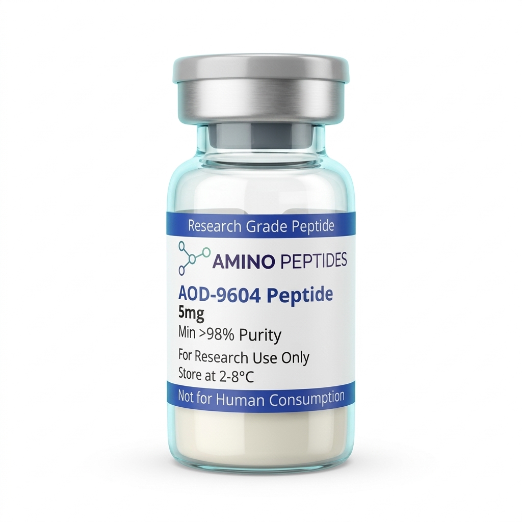
AOD-9604 Peptide
From £22
Supplied as a sealed freeze-dried vial (stable 12–24 months at 2°C–8°C in a standard refrigerator).
Shop AOD-9604 Peptide →Instead, the aod9064 peptide focuses its activity on beta-3 adrenergic receptors sitting on the surface of white adipocytes (fat cells). In obese animal models, the density of these beta-3 receptors often drops, making it hard for the cells to release stored triglycerides. Published studies observed that introducing AOD-9604 brought beta-3 receptor expression back up to the levels seen in lean control animals. At the same time, the fragment works in two directions inside the fat cell: it stimulates lipolysis (the cleaving of stored fat into free fatty acids for cellular energy) and slows down lipogenesis (the creation of new fatty deposits from dietary carbohydrates or lipids). By zeroing in on fat cell metabolism without touching the glucose-regulating machinery, this fragment gives researchers a clean way to study lipid mobilisation in isolation.
What Do Preclinical Timelines Show From Day One to Week Twelve?
In laboratory settings, the biological response to AOD-9604 unfolds in two distinct phases: an immediate biochemical shift that happens within hours, followed by gradual structural changes in adipose reserves that take several weeks to measure. Understanding this split helps researchers set realistic observation windows when designing metabolic assays.
During the first 24 to 72 hours of a study, the action is purely chemical and happens at the cell membrane and inside the cytosol. Shortly after exposure in isolated adipocyte cultures or live rodent models, researchers can measure a rapid uptick in glycerol and free fatty acid release into the surrounding medium or bloodstream. This early spike is the direct fingerprint of lipolysis. Simultaneously, respiratory quotient measurements in metabolic cages begin to dip within the first few days. A lower respiratory quotient tells investigators that the test subjects are oxidising a higher ratio of lipids for cellular energy rather than burning strictly carbohydrates. Throughout this early window, blood glucose monitors and serum insulin assays remain completely steady, confirming that the acute fat-releasing signal does not trigger a glycemic bounce.
Between weeks two and four, genetic and enzymatic shifts become visible in tissue samples. Lab assays from this middle phase show increased messenger RNA (mRNA) for beta-3 adrenergic receptors in white adipose depots, alongside a noticeable drop in the activity of lipogenic enzymes like fatty acid synthase and acetyl-CoA carboxylase. In simple terms, the fat cells are not only releasing stored energy faster, they are also turning down their internal fat-storage factories. Even so, gross changes in total body mass during the first fortnight are usually modest in rodent models, as water balance and cellular remodelling take time to settle.
The macroscopic structural changes typically show up between weeks four and twelve in published studies. In genetically obese mice (such as ob/ob models) observed over a twelve-week span, cumulative reductions in white adipose tissue weight become clear when compared against untreated controls. Interestingly, preclinical models demonstrated that lean animals with low baseline body fat showed very little change in mass when exposed to the same measured levels, suggesting the fragment relies on the presence of excess lipid stores and suppressed beta-3 receptors to show its full effect. Separate cartilage and joint tissue remodelling studies in rabbit models also tracked structural recovery over four- to eight-week timelines, where proteoglycan and collagen synthesis gradually improved without triggering abnormal bone overgrowth.
Reconstitution calculator
Choose a vial mass and a bacteriostatic reconstitution solution volume. The figure is the resulting concentration.
Concentration: 2.50 mg/mL
Per 0.01mL (10µL): 25.0 mcg
Open the full calculatorBacteriostatic reconstitution solutionAOD-9604 vs Full-Length hGH vs Unmodified Fragment 176-191 in Laboratory Models
| Characteristic | AOD-9604 (Tyr-hGH 177-191) | Full-Length hGH (191 AA) | Unmodified Fragment 176-191 |
|---|---|---|---|
| Amino Acid Length | 16 amino acids (N-terminal tyrosine added to residues 177-191) | 191 amino acids (complete polypeptide chain) | 16 amino acids (native residues 176-191) |
| Primary Receptor Binding | Does not bind to the canonical hGH receptor; acts on adipose beta-3 adrenergic pathways | High affinity for transmembrane hGH receptors across all tissues | Negligible affinity for canonical hGH receptors; targets adipose signalling |
| Effect on IGF-1 Levels | No observed change in circulating IGF-1 concentrations | Strongly upregulates hepatic and local IGF-1 production | No observed change in circulating IGF-1 concentrations |
| Carbohydrate and Insulin Impact | Does not induce insulin resistance or alter blood glucose levels in laboratory models | Can reduce insulin sensitivity and elevate blood glucose concentrations | Maintains normal glucose tolerance and insulin signalling in preclinical models |
| Lipolytic Mechanism | Stimulates lipolysis and inhibits lipogenesis; restores beta-3 adrenergic receptor expression | Stimulates lipolysis alongside broad anabolic and mitogenic tissue proliferation | Stimulates lipolysis and suppresses fatty acid synthase activity in adipose tissue |
Which Complementary Sequences Pair With AOD-9604 in Metabolic Research?
Because AOD-9604 operates through a very narrow pathway—targeting fat cell beta-3 receptors and lipid enzymes without raising IGF-1 or shifting blood sugar—researchers frequently pair it with other compounds to study multi-angle metabolic regulation or tissue recovery. When scientists explore peptide hormones for energy balance, combining sequences that hit separate cellular targets avoids overwhelming a single receptor type.
One common pairing in metabolic laboratory models involves combining AOD-9604 with MOTS-c, a sixteen-amino-acid mitochondrial-derived peptide. These two molecules handle opposite ends of the cellular energy pipeline. First, AOD-9604 encourages white adipocytes to break down stored triglycerides and release free fatty acids into circulation. However, liberating fatty acids is only half the puzzle; those lipids still need to be burned inside the mitochondria. That is where MOTS-c enters the picture. Published studies show that MOTS-c activates the AMPK pathway and helps shuttle both glucose and fatty acids into the mitochondrial furnace for beta-oxidation. Studying them side by side allows investigators to watch both the supply phase (lipid release via AOD-9604) and the demand phase (mitochondrial fuel burning via MOTS-c) without overlap.
Another frequent comparison and complementary pairing involves 5-Amino-1MQ, a small molecule that inhibits the enzyme nicotinamide N-methyltransferase (NNMT). High NNMT levels in fat tissue slow down the nicotinamide adenine dinucleotide (NAD+) salvage pathway, which sluggishly drags down cellular metabolism. While AOD-9604 works on surface adrenergic signalling and lipogenic enzymes, 5-Amino-1MQ boosts intracellular NAD+ levels and increases the expression of GLUT4 transporters. In experimental designs, pairing an extracellular lipid-mobiliser with an intracellular NAD+ booster lets researchers observe how fat cells behave when both membrane signalling and internal coenzyme pools are supported at once.
Outside of pure lipid research, AOD-9604 has also drawn interest in structural tissue remodelling stacks alongside BPC-157. A lesser-known series of preclinical models in rabbits looked at how AOD-9604 encouraged chondrocyte (cartilage cell) proliferation and matrix production in worn joints. Because BPC-157 supports fibroblast migration and microvascular blood flow in tendons and ligaments, studying the two together gives researchers a way to observe soft-tissue vascular recovery alongside cartilage matrix synthesis. In every one of these combinations, the appeal of AOD-9604 is that it never brings unwanted insulin spikes or IGF-1 elevation into the experiment, keeping the biochemical background clean.
Figure: AOD-9604: How This Fragment Targets Adipose Tissue Without Affecting Blood Sugar
Why Disulphide Bridging, TFA Removal, and Cold Storage Dictate Vial Reliability
When working with a sequence like AOD-9604, the reliability of any laboratory observation depends heavily on how the peptide was synthesised, folded, and purified before it ever reaches the vial. Unlike simple linear peptides, AOD-9604 contains two cysteine residues at positions 182 and 189 that must form a precise internal disulphide bridge. This tiny sulphur-to-sulphur bond pulls the amino acid chain into a cyclic loop. If a synthesis facility cuts corners and leaves those cysteines unbonded—or worse, allows separate peptide chains to link together into tangled dimers—the molecule loses its three-dimensional shape and will not fit the target receptors on fat cells.
High-Performance Liquid Chromatography (HPLC) testing at 99% purity or higher is the only way to confirm that the cyclic loop formed properly and that truncated failure sequences were washed away. Alongside HPLC, Mass Spectrometry (MS) verifies the exact molecular weight of 1815.1 daltons, proving that the N-terminal tyrosine cap is in place and the disulphide bridge is closed. Sourcing vials accompanied by batch-specific chromatograms removes the risk of running a six-week metabolic assay with inactive, misfolded chains.
Just as important is the removal of trifluoroacetic acid (TFA) salts left behind during solid-phase synthesis. Cheaply processed peptides often carry a heavy load of residual TFA. In delicate adipocyte cultures, free TFA alters the pH of the medium, stresses cell membranes, and causes erratic signalling readouts that look like metabolic shifts but are actually just chemical irritation. Premium research preparations swap out harsh TFA for a mild acetate counter-ion or lyophilise the batch to ultra-low salt levels, keeping the cellular environment calm and predictable.
Once the lyophilised powder arrives in the laboratory, proper cold-chain handling preserves that delicate cyclic ring. Unreconstituted vials can be kept at minus 20°C for long-term storage, while active working vials belong in a dedicated laboratory refrigerator held between 2°C and 8°C. When preparing the peptide for an assay, adding a sterile bacteriostatic reconstitution solution slowly down the inside glass wall prevents foaming. Never shake the vial vigorously; mechanical shearing can strain the disulphide bridge and cause the hydrophobic tyrosine tail to clump together, turning a clear solution cloudy. Gentle swirling and strict 2°C to 8°C fridge storage ensure the fragment stays intact from the first day of the protocol to the last.
Frequently Asked Questions
Why do researchers study AOD-9604 for fat metabolism without blood sugar changes?
Researchers examine AOD-9604 because it isolates the fat-reducing section of the growth hormone molecule while leaving out the regions that alter insulin or glucose levels. In published studies using obese rodent models, this specific sixteen-amino-acid fragment stimulated fat breakdown and slowed fat storage without causing hyperglycaemia or unwanted cartilage growth.
What timeline do preclinical models show for AOD-9604 observations?
Preclinical models typically show measurable shifts in fat oxidation within two to four weeks of daily administration. During twelve-week laboratory evaluations, scientists observed steady reductions in adipose mass alongside improved beta-3 adrenergic receptor expression. The exact timeline depends heavily on the measured level used and the specific metabolic baseline of the animal model.
How is AOD-9604 combined with other compounds in laboratory protocols?
In laboratory settings, scientists occasionally pair AOD-9604 with other metabolic fragments or growth hormone secretagogues to observe combined signalling pathways. However, most foundational researchers prefer testing AOD-9604 alone to clearly characterise its isolated effect on adipose tissue. Mixing compounds requires careful protocol ratios to ensure one peptide does not cloud the data.
How should AOD-9604 purity be verified and stored in the fridge?
Researchers verify AOD-9604 purity by checking third-party High-Performance Liquid Chromatography certificates that confirm a purity level above ninety-eight percent. Unmixed lyophilised powder stays stable in a freezer at minus twenty degrees Celsius. Once mixed with a bacteriostatic reconstitution solution, the liquid vial must stay refrigerated between two and eight degrees Celsius.
Bibliography
- Ng FM, Sun J, Sharma L, Libinaka R, Jiang WJ, Gianello R. Metabolic studies of a synthetic lipolytic domain (AOD9604) of human growth hormone. Horm Res. 2000;53(6):274-278. View published research
- Heffernan M, Summers RJ, Thorburn A, Ogru E, Gianello R, Jiang WJ, Ng FM. The effects of human GH and its lipolytic fragment (AOD9604) on lipid metabolism following chronic protocol exposure in obese mice and beta(3)-AR knock-out mice. Endocrinology. 2001;142(12):5182-5189. View published research
- Heffernan MA, Thorburn AW, Fam B, Summers R, Conway-Campbell B, Waters MJ, Ng FM. Increase of fat oxidation and weight loss in obese mice caused by chronic exposure to human growth hormone or a modified C-terminal fragment. Int J Obes Relat Metab Disord. 2001;25(10):1442-1449. View published research
- Wu Z, Ng FM. Antilipogenic action of synthetic C-terminal sequence 177-191 of human growth hormone. Biochem Mol Biol Int. 1993;30(1):187-196. View published research
⚠️ Research Use Only Disclaimer: All peptides and compounds are sold strictly for in-vitro laboratory research purposes only. Not intended for human or veterinary use, not a dietary supplement, and not approved to diagnose, treat, cure, or prevent any disease or condition. For use by qualified researchers in suitably equipped laboratory environments only. Amino Peptides Ltd operates in full compliance with UK MHRA regulations and applicable EU research chemical guidelines.