
Assessing the Efficacy of KPV Peptide in Modulating In-Vitro Inflammatory Pathways
30th Sep 2026

What You Need to Know About KPV Peptide
- Researchers frequently investigate KPV to understand how tiny protein fragments can calm overactive immune responses without altering skin pigmentation.
- In plain terms, KPV enters stressed cells via specialized peptide transporters and blocks the master switch responsible for producing inflammatory alarm signals.
- Published studies in cell cultures demonstrate that KPV lowers measured levels of pro-inflammatory messengers like TNF-alpha and interleukin-6.
- Preclinical models show that this three-amino-acid sequence supports epithelial tissue remodelling and cellular recovery through PepT1-mediated uptake.
How a Three-Amino-Acid Fragment Quiets Cellular Stress Signals
KPV calms overactive immune signalling in cell cultures by entering the cell and blocking the NF-kB pathway before inflammatory messengers can be switched on. Because it consists of only three amino acids—lysine, proline, and valine—it achieves this calming effect without triggering the pigment-changing receptors linked to its larger parent hormone.
To understand how this tiny tripeptide works, it helps to look at where it comes from. KPV is the tail-end fragment of alpha-melanocyte-stimulating hormone (alpha-MSH). While the full hormone binds to surface receptors that control skin and hair darkening, published studies show that snipping off just the last three amino acids leaves behind a compact molecule that focuses purely on cellular recovery and stress control.
When Assessing the Efficacy of KPV Peptide in Modulating In-Vitro Inflammatory Pathways, researchers focus heavily on a protein complex called nuclear factor kappa B (NF-kB). Think of NF-kB as a cellular smoke alarm. When epithelial or immune cells meet an irritant like bacterial lipopolysaccharide (LPS), an inhibitory protein called I-kappa-B breaks down. This frees NF-kB to travel into the cell nucleus, where it instructs the DNA to pump out pro-inflammatory cytokines such as TNF-alpha, IL-6, and IL-8.
Published studies demonstrate that KPV steps in right at this nuclear doorway. Instead of merely sitting on the outside of the cell membrane, KPV hitches a ride inside through a specialised protein doorway known as the PepT1 transporter. Once inside the cytoplasm, it stabilises I-kappa-B and physically competes with NF-kB for entry into the nucleus through importin proteins. With the doorway blocked, the cell stops broadcasting stress signals, allowing local tissue recovery to proceed without runaway swelling or oxidative damage.
Because PepT1 transporters are heavily expressed in stressed gut lining cells and keratinocytes, preclinical models show that KPV naturally concentrates right where cellular irritation is highest. This targeted uptake explains why such a small molecule produces measurable drops in cytokine output across diverse laboratory models without shutting down normal, everyday cellular housekeeping.
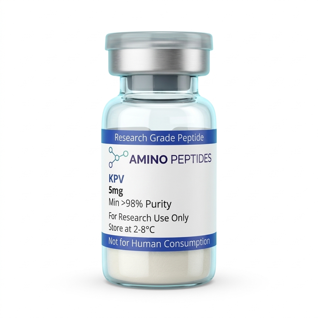
KPV Peptide
From £25
Supplied as a sealed freeze-dried vial (stable 12–24 months at 2°C–8°C in a standard refrigerator).
Shop KPV Peptide →What Cell Culture Clocks Show From First Contact to Tissue Remodelling
Watching KPV work in a petri dish requires looking at two distinct phases: the rapid biochemical quietening that happens in the first few hours, and the slower structural recovery of the cell layer that unfolds over several days. Laboratory models tracking these changes use specific time points to measure how quickly the peptide enters the cell and when physical barrier repair actually finishes.
During the first fifteen to sixty minutes after introducing a measured level of KPV into a stressed cell culture, the main event is transport and nuclear blocking. In cultured human colonic epithelial cells (such as Caco-2 lines) exposed to inflammatory triggers, PepT1 transporters pull the tripeptide across the membrane within minutes. By the two-hour mark, Western blot assays typically record a clear drop in phosphorylated NF-kB inside the nucleus compared to untreated control wells. At this early stage, the cells look no different under a microscope, but their internal chemical signalling has already shifted away from alarm mode.
Between six and twenty-four hours, the downstream effects of that quietened nucleus become easy to measure in the liquid surrounding the cells. ELISA tests run at the twelve-hour mark consistently show lower concentrations of secreted IL-8 and TNF-alpha. Because the cells are no longer spending their ATP reserves manufacturing inflammatory proteins, cellular energy shifts back toward basic maintenance and survival.
Structural tissue remodelling takes longer and generally shows up between forty-eight and ninety-six hours in vitro. Researchers often measure this phase using transepithelial electrical resistance (TEER), which tests how tightly cells are zipped together in a flat sheet, alongside scratch-wound closure assays. By day three or four, cell layers given a steady protocol ratio of KPV rebuild their tight-junction proteins—like occludin and zonula occludens-1—much faster than irritated control cultures. The physical gaps between cells close up, restoring a solid barrier against outside irritants.
Comparison of KPV and Full-Length Alpha-MSH in In-Vitro Laboratory Models
| Parameter | KPV Tripeptide (Lys-Pro-Val) | Full-Length Alpha-MSH (13 Amino Acids) |
|---|---|---|
| Amino Acid Length | Three residues (residues 11 to 13 of alpha-MSH) | Thirteen residues (intact parent hormone) |
| Primary Cellular Uptake Route | PepT1 di- and tripeptide membrane transporter | Melanocortin surface receptors (MC1R, MC3R, MC4R, MC5R) |
| Pigmentation Signalling | Does not trigger melanin synthesis in melanocyte assays | Strongly activates MC1R to stimulate melanin production |
| NF-kappaB Pathway Modulation | Inhibits p65 nuclear translocation and stabilizes I-kappaB-alpha | Suppresses NF-kappaB via cyclic AMP (cAMP) receptor cascades |
| Epithelial Barrier Observations | Reduces cytokine output in intestinal and bronchial cell lines | Broad systemic signalling across multiple tissue types |
Pairing KPV With Structural and Barrier-Support Peptides in the Lab
Because KPV focuses so specifically on intracellular NF-kB silencing, researchers frequently combine it with other peptides that handle different stages of tissue recovery. In laboratory models, pairing an internal signalling brake with molecules that build extracellular matrix or defend against microbes lets investigators watch multiple repair pathways run at the same time without overlapping or competing for the same receptors.
A frequent pairing in barrier-defence research involves KPV and LL-37. While KPV slips inside the epithelial cell via PepT1 to quieten excessive cytokine release, the cathelicidin peptide LL-37 works out in the extracellular space to disrupt bacterial membranes and neutralise endotoxins like LPS. Studying them side by side in co-culture assays allows scientists to observe how neutralising outside triggers while simultaneously calming internal nuclear signalling protects fragile cell sheets from breaking down under heavy microbial stress.
Another widely studied approach pairs KPV with structural recovery peptides like BPC-157, TB-500 (Thymosin Beta-4 fragment), and the copper-binding tripeptide GHK-Cu. In multi-pathway laboratory protocols, researchers often source a pre-formulated klow blend to examine how these four distinct mechanisms interact within the same culture environment. Each component handles a separate job during tissue remodelling.
In this four-part setup, KPV keeps the local inflammatory signalling in check during the early hours so that freshly dividing cells are not damaged by oxidative stress. Meanwhile, BPC-157 upregulates growth hormone receptor expression on fibroblasts and encourages nitric oxide balance, and TB-500 binds to actin to help cells migrate across physical gaps. Finally, GHK-Cu supplies bioavailable copper to enzymes like lysyl oxidase, which cross-links new collagen and elastin fibres during the later structural phase. Combining all four in controlled preclinical models shows how calming the cellular environment first makes subsequent structural rebuilding far more organised.

Figure: Assessing the Efficacy of KPV Peptide in Modulating In-Vitro Inflammatory Pathways
Reconstitution calculator
Choose a vial mass and a bacteriostatic reconstitution solution volume. The figure is the resulting concentration.
Concentration: 2.50 mg/mL
Per 0.01mL (10µL): 25.0 mcg
Open the full calculatorBacteriostatic reconstitution solutionWhy Salt Removal and Cold Storage Decide Your Assay Results
When working with a tiny tripeptide in sensitive cell cultures, the cleanliness of the lyophilized powder directly decides whether an experiment succeeds or fails. Because Assessing the Efficacy of KPV Peptide in Modulating In-Vitro Inflammatory Pathways relies on measuring tiny changes in cytokine output, any leftover manufacturing chemicals inside the vial can irritate the cells and ruin the readout.
The biggest hidden culprit in low-grade peptide vials is trifluoroacetic acid (TFA). TFA is a harsh acid used during solid-phase synthesis to cleave the finished peptide chain from its resin bead. If a manufacturer skips proper ion-exchange chromatography, residual TFA stays bound to the lysine amino acid in KPV as a salt. When you dissolve a TFA-heavy powder and add it to living epithelial cells, the acid lowers the local pH and causes cellular stress all on its own. This triggers the exact NF-kB inflammation you are trying to calm, creating confusing data where the peptide appears weaker than it truly is. Choosing vials verified at 99% or higher purity via High-Performance Liquid Chromatography (HPLC) with confirmed TFA removal prevents this chemical interference.
Once a clean, mass-spectrometry-verified vial arrives in the lab, temperature control keeps the three amino acids intact. In its dry, freeze-dried state, KPV is sturdy, but it should still be stored in a dedicated refrigerator between 2°C and 8°C away from bright light. Before opening the vial, letting it warm up to room temperature inside a desiccator stops airborne moisture from condensing onto the dry cake.
To prepare the peptide for an assay, gently trickle bacteriostatic reconstitution solution down the inside glass wall rather than squirting it straight onto the powder. Never shake the vial; a gentle swirl dissolves the tripeptide in seconds without shearing or foaming. Once mixed into liquid form, keep the vial refrigerated at 2°C to 8°C and avoid repeated freeze-thaw cycles so every measured level added to your culture wells performs identically from day one to the end of the study.
Frequently Asked Questions
Why are researchers assessing the efficacy of KPV peptide in modulating in-vitro inflammatory pathways?
Researchers study KPV because this tiny three-amino-acid fragment calms overactive immune signalling in laboratory cell cultures without triggering unwanted pigmentation changes. Published studies show it enters cells to quieten the NF-kappaB pathway, helping scientists understand how tissue recovery happens at a microscopic level.
How quickly does KPV alter inflammatory markers in laboratory cell models?
In laboratory models, KPV begins quieting inflammatory signalling within just a few hours of exposure. Scientists typically measure significant drops in pro-inflammatory proteins between six and twenty-four hours after introducing a specific study amount, while broader cellular barrier recovery observations often run across several days.
Do laboratory studies combine KPV with other peptides like BPC-157?
Yes, preclinical models frequently pair KPV with compounds such as BPC-157 or TB-500 to observe combined effects on tissue remodelling. Whilst KPV focuses on calming inflammatory signalling inside the cell, BPC-157 is analysed for supporting structural cell migration, allowing researchers to study multiple recovery pathways simultaneously.
What purity level and storage conditions are required for KPV in the laboratory?
Laboratory studies require KPV at a minimum of ninety-eight percent purity, verified by HPLC and mass spectrometry, to prevent contaminants from skewing cell assays. Lyophilised powder is stored frozen at minus twenty degrees Celsius, whilst any mixture prepared with bacteriostatic reconstitution solution is kept refrigerated between two and eight degrees.
Bibliography
- Dalmasso G, Charrier-Hisamuddin L, Nguyen HT, et al. PepT1-mediated tripeptide KPV uptake reduces intestinal inflammation. Gastroenterology. 2008;134(1):166-178. View published research
- Land SC. Inhibition of cellular and systemic inflammation cues in human bronchial epithelial cells by melanocortin-related peptides: mechanism of KPV action and a role for MC3R agonists. Int J Physiol Pathophysiol Pharmacol. 2012;4(2):59-73. View published research
- Brzoska T, Luger TA, Maaser C, Abels C, Bohm M. Alpha-melanocyte-stimulating hormone and related tripeptides: biochemistry, antiinflammatory and protective effects in vitro and in vivo, and future perspectives for the protocol of immune-mediated inflammatory diseases. Endocr Rev. 2008;29(5):581-602. View published research
- Kannengiesser K, Maaser C, Heidemann J, et al. Melanocortin-derived tripeptide KPV has anti-inflammatory potential in murine models of inflammatory bowel disease. Inflamm Bowel Dis. 2008;14(3):324-331. View published research
⚠️ Research Use Only Disclaimer: All peptides and compounds are sold strictly for in-vitro laboratory research purposes only. Not intended for human or veterinary use, not a dietary supplement, and not approved to diagnose, treat, cure, or prevent any disease or condition. For use by qualified researchers in suitably equipped laboratory environments only. Amino Peptides Ltd operates in full compliance with UK MHRA regulations and applicable EU research chemical guidelines.