
The Bioenergetic Trio: Stacking NAD+, MOTS-c, and SS-31 for Cellular Research
11th Sep 2026

What You Need to Know About NAD+, MOTS-c & SS-31
- People look this stack up because the compounds touch cellular energy from different directions.
- NAD+ feeds redox reactions. MOTS-c is a mitochondrial peptide. SS-31 is discussed for the inner membrane when it is part of the stack.
- Published work tracks early signalling within hours and later changes in mitochondrial output.
- A 99%+ HPLC-verified, freeze-dried vial kept at 2°C–8°C is the variable a laboratory can actually hold constant.
How NAD+, MOTS-c, and SS-31 Power Up Cellular Energy Models
In laboratory models, combining NAD+, MOTS-c, and SS-31 targets three separate bottlenecks in mitochondrial adenosine triphosphate (ATP) production at the same time. SS-31 stabilises the inner mitochondrial membrane, NAD+ supplies the raw electron carrier needed for energy conversion, and MOTS-c signals the cell to take up and burn fuel more efficiently.
To see why researchers study these three together, it helps to picture a mitochondrion as a small power station. Inside that power station, electrons pass along a chain of proteins set into an inner wall. That inner wall is rich in a special fat molecule called cardiolipin. When a cell faces oxidative stress or ageing in a dish, cardiolipin breaks down, the inner wall loses its tight folds, and electrons leak out as damaging reactive oxygen species instead of making ATP.
SS-31, also known in literature as elamipretide, is a tiny four-amino-acid peptide that heads straight to that inner wall. Published studies show it binds directly to cardiolipin, holding the tight folds together so the electron transport chain can run without leaking sparks. Once the physical structure is steady, the system needs fuel carriers. That is where nicotinamide adenine dinucleotide (NAD+) enters the picture. NAD+ catches high-energy electrons from broken-down sugars and fats, becoming NADH, and drops those electrons straight into the first station of the transport chain.
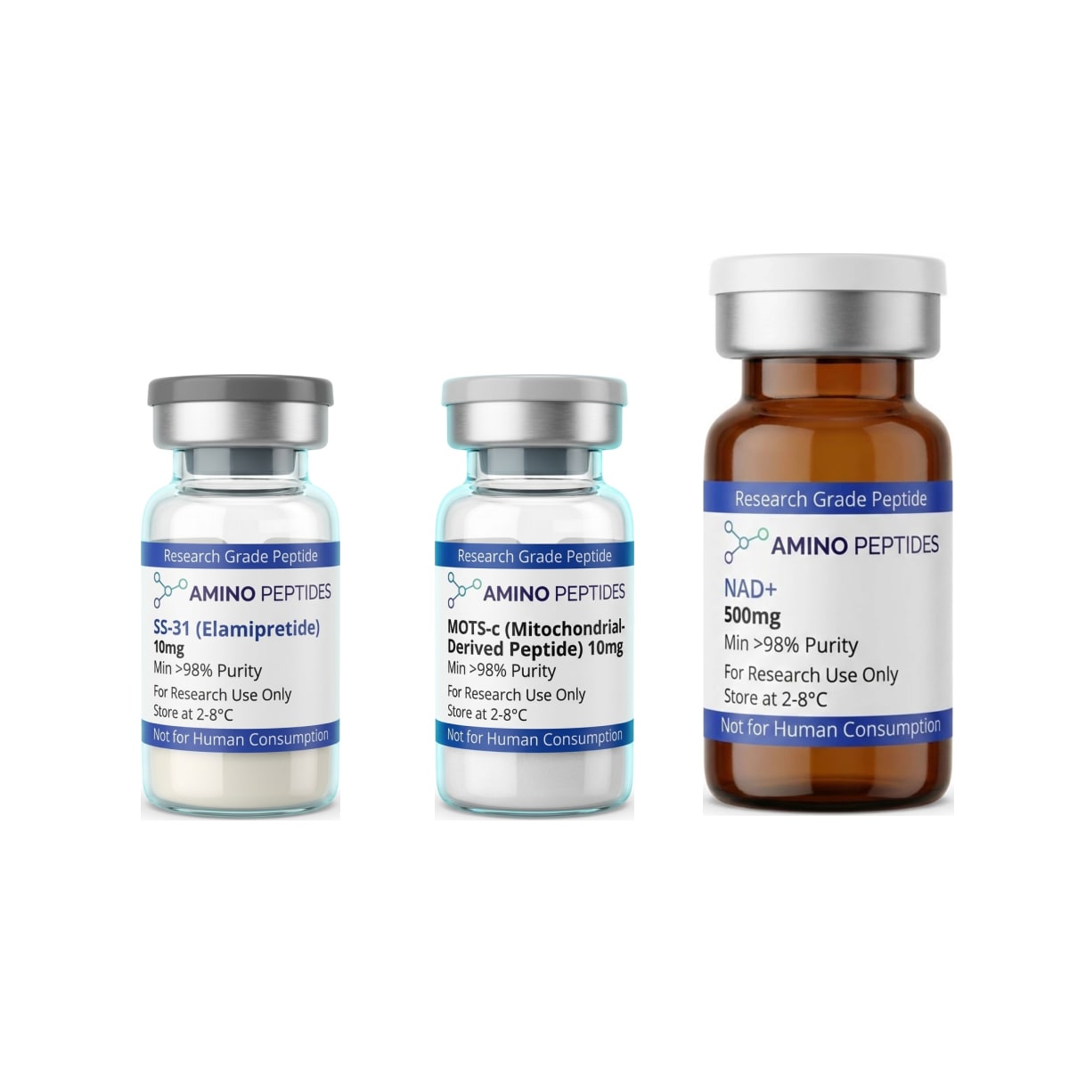
SS-31 & MOTS-c & NAD+
From £116
Supplied as a sealed freeze-dried vial (stable 12–24 months at 2°C–8°C in a standard refrigerator).
Shop SS-31 & MOTS-c & NAD+ →Even with a steady wall and plenty of NAD+, a cell still needs a manager to regulate how much fuel enters the system. MOTS-c is a sixteen-amino-acid peptide encoded by the mitochondrial DNA itself. Under metabolic stress, MOTS-c moves to the cell nucleus and activates the AMPK signalling pathway. This tells the cell to pull in more glucose and fatty acids for clean burning. Together, the three compounds cover structural repair, electron transport, and metabolic signalling in one coordinated experimental setup.
What Laboratory Timelines Show From First Signal to Membrane Repair
When investigators introduce NAD+, MOTS-c, and SS-31 into preclinical models, the observed cellular responses do not happen all at once. Instead, published studies describe a clear sequence that moves from rapid chemical signalling in the first few hours to physical tissue remodelling over several weeks.
The earliest changes appear within minutes to hours and are led by SS-31 and NAD+. Because SS-31 easily crosses the outer cell membrane and concentrates inside the mitochondria by several hundred-fold, laboratory assays often record a drop in reactive oxygen species and a jump in oxygen consumption within the first one to six hours. During this same early window, adding exogenous NAD+ or its immediate precursors raises the intracellular NAD+ to NADH ratio. This rapid shift switches on sirtuin enzymes, especially SIRT1 and SIRT3, which begin deacetylating mitochondrial proteins to optimise their speed.
The middle phase takes place between twenty-four and seventy-two hours. Here, the gene expression shifts triggered by MOTS-c become easy to measure. Once MOTS-c activates the AICAR-AMPK pathway and moves into the nucleus, it alters transcription factors that govern stress resistance and glucose uptake. Researchers measuring cell cultures at the two-day mark consistently note higher levels of GLUT4 transporters on cell surfaces and improved clearance of damaged proteins.
Longer structural changes require days to weeks of sustained protocol ratios in animal or three-dimensional tissue models. Over a two-to-four-week observation period, electron microscopy in published studies reveals actual physical regeneration of the mitochondrial cristae — the folded inner membranes looked fresh and tightly packed again rather than swollen and fragmented. At this later stage, researchers also measure mitochondrial biogenesis, meaning the cells have actually built brand-new mitochondria through PGC-1alpha signalling, shifting the tissue toward a younger, more energetic baseline profile.
Reconstitution calculator
Choose a vial mass and a bacteriostatic reconstitution solution volume. The figure is the resulting concentration.
Concentration: 2.50 mg/mL
Per 0.01mL (10µL): 25.0 mcg
Open the full calculatorBacteriostatic reconstitution solutionBiochemical Comparison of the Bioenergetic Trio in Preclinical Research
| Research Parameter | NAD+ (Nicotinamide Adenine Dinucleotide) | MOTS-c (Mitochondrial Open Reading Frame) | SS-31 (Elamipretide) |
|---|---|---|---|
| Molecular Class | Dinucleotide coenzyme (663.43 g/mol) | 16-amino-acid polypeptide (2174.6 g/mol) | Synthetic tetrapeptide (639.8 g/mol) |
| Primary Cellular Target | Sirtuin enzymes (SIRT1-7), PARPs, and redox reactions | Folate-methionine cycle and AMPK signalling pathway | Cardiolipin on the inner mitochondrial membrane |
| Core Mechanism | Transfers electrons in the Krebs cycle and electron transport chain | Translocates to the nucleus during stress to regulate metabolic genes | Binds cardiolipin to optimise cristae curvature and supercomplex stability |
| Observed Preclinical Outcome | Restores redox balance and supports DNA repair enzymes in aged cell lines | Improves glucose clearance and metabolic flexibility in rodent models | Reduces reactive oxygen species leakage and preserves ATP synthesis |
| Role in the Trio Stack | Provides the raw cofactor fuel required for oxidative phosphorylation | Acts as the metabolic software signalling the cell to utilise nutrients | Maintains the physical hardware of the mitochondrial electron transport chain |
Building Smarter Mitochondrial Stacks With Complementary Research Peptides
While pairing NAD+, MOTS-c, and SS-31 covers the main steps of ATP generation, researchers often look at how this core group interacts with broader signalling pathways. Depending on whether an experiment focuses on metabolic turnover, cellular cleanup, or endocrine signalling, scientists frequently pair these compounds with a pre-formulated mitoprime peptide research stack to keep their baseline variables consistent across multiple assay plates.
One common area of investigation combines mitochondrial repair agents with compounds that alter fatty acid mobilisation. For instance, MOTS-c naturally pushes cells to oxidise fats through the AMPK pathway. In adipocyte and skeletal muscle studies, researchers sometimes examine how this interacts with lipolytic fragments like AOD-9604 or 5-Amino-1MQ. While 5-Amino-1MQ blocks the NNMT enzyme to preserve natural NAD+ levels inside the cytosol, MOTS-c and SS-31 make sure the mitochondria are ready to burn the resulting fatty acids cleanly without creating excess free radical damage.
Another avenue of laboratory inquiry looks at how mitochondrial energy availability affects wider systemic messengers, including peptide hormones for endocrine and receptor-binding studies. Producing and secreting signalling peptides takes a massive amount of cellular ATP. When researchers study growth hormone secretagogues such as Ipamorelin or CJC-1295 alongside mitochondrial support compounds, they can observe whether keeping cellular energy high prevents receptor fatigue during extended observation periods.
Finally, investigators studying cellular senescence often alternate an energy-building phase with an autophagic cleanup phase. In these protocols, peptides like Epitalon or FOXO4-DRI are studied alongside NAD+ and SS-31. The logic observed in preclinical models is straightforward: NAD+ and SS-31 give healthy cells the ATP they need to carry out routine maintenance, while the longevity-focused peptides help clear out senescent cells that can no longer be repaired.

Figure: The Bioenergetic Trio: Stacking NAD+, MOTS-c, and SS-31 for Cellular Research
Why HPLC Verification and Cold Storage Protect Your Experimental Data
Running multi-compound experiments with NAD+, MOTS-c, and SS-31 means every single reagent must be free of hidden chemical noise. If even one vial in a three-part protocol contains synthesis leftovers or broken peptide fragments, it becomes impossible to tell which molecule caused an observed cellular shift and which one triggered a stress response.
The most common contaminant in poorly finished peptides is trifluoroacetic acid (TFA). TFA is a strong acid used during solid-phase synthesis to cleave the finished peptide chain from its resin bead. If a manufacturer skips the extra ion-exchange steps needed to remove TFA salts, that residual acid stays in the lyophilised powder. When you add your liquid carrier and place the solution onto delicate cell cultures, excess TFA drops the local pH and damages mitochondrial membranes on contact. That directly fights against the membrane-stabilising work of SS-31. Sourcing vials verified at 99% or higher purity via High-Performance Liquid Chromatography (HPLC) and Mass Spectrometry ensures those harsh synthesis byproducts have been scrubbed out.
Storage and reconstitution habits matter just as much as the initial certificate of analysis. Both MOTS-c and SS-31 rely on intact amino acid sequences that begin to break down if left at warm room temperatures after mixing. In dry, lyophilised form, vials stay stable in a freezer at minus 20°C. Once mixed with a sterile bacteriostatic reconstitution solution, the vials must live in a standard laboratory refrigerator kept strictly between 2°C and 8°C.
NAD+ requires extra care during mixing because it is a coenzyme rather than a simple peptide chain, and its molecular structure is sensitive to extreme pH swings. Always let refrigerated vials warm up to room temperature for ten minutes before opening them so condensation does not ruin the powder, and swirl the liquid gently down the glass wall instead of shaking the vial.
Frequently Asked Questions
Why are researchers searching for the NAD+, MOTS-c, and SS-31 trio?
Researchers investigate this trio because each compound targets a distinct step of mitochondrial energy production in laboratory models. Published studies show SS-31 stabilises the inner membrane, NAD+ drives electron transport, and MOTS-c optimises metabolic signalling. Together, they help scientists analyse complete cellular recovery and energy pathways during age-related decline.
What is a typical study timeline for observing this bioenergetic stack?
In preclinical models, measurable shifts in cellular energy typically appear within two to four weeks of consistent observation. Short-term laboratory protocols track immediate ATP output and membrane stability within days. Longer eight-week investigations are generally required to characterise lasting tissue remodelling, gene expression changes, and full metabolic adaptation.
Can NAD+, MOTS-c, and SS-31 be combined in the same laboratory protocol?
Yes, researchers frequently pair these three compounds in laboratory models to examine complementary mitochondrial pathways without overlapping receptor fatigue. Rather than mixing them in a single vial, protocols usually apply each compound separately at its specific study amount. This approach keeps each peptide stable and clarifies individual signalling effects.
How should these research compounds be verified for purity and stored?
Every batch should exceed ninety-nine percent purity as verified by independent High-Performance Liquid Chromatography and Mass Spectrometry testing. Lyophilised powders remain stable in a freezer at minus twenty degrees Celsius. Once mixed with bacteriostatic reconstitution solution, vials must stay refrigerated between two and eight degrees Celsius away from light.
Bibliography
- Covarrubias AJ, Perrone R, Grozio A, Verdin E. NAD+ metabolism and its roles in cellular processes during ageing. Nature Reviews Molecular Cell Biology. 2021;22(2):119-141. View published research
- Lee C, Zeng J, Drew BG, et al. The mitochondrial-derived peptide MOTS-c promotes metabolic homeostasis and reduces obesity and insulin resistance. Cell Metabolism. 2015;21(3):443-454. View published research
- Szeto HH. First-in-class cardiolipin-protective compound as a biological agent to restore mitochondrial bioenergetics. British Journal of Pharmacology. 2014;171(8):2029-2050. View published research
- Chavez JD, Tang X, Campbell MD, et al. Mitochondrial protein interaction landscape of SS-31. Proceedings of the National Academy of Sciences. 2020;117(26):15363-15373. View published research
- Kim KH, Son JM, Benayoun BA, Lee C. The Mitochondrial-Encoded Peptide MOTS-c Translocates to the Nucleus to Regulate Nuclear Gene Expression in Response to Metabolic Stress. Cell Metabolism. 2018;28(3):516-524. View published research
⚠️ Research Use Only Disclaimer: All peptides and compounds are sold strictly for in-vitro laboratory research purposes only. Not intended for human or veterinary use, not a dietary supplement, and not approved to diagnose, treat, cure, or prevent any disease or condition. For use by qualified researchers in suitably equipped laboratory environments only. Amino Peptides Ltd operates in full compliance with UK MHRA regulations and applicable EU research chemical guidelines.