
Characterisation of Bioluminescent Glow Kinetics in Microplate-Based Luciferase Reporter Assays
16th Nov 2026
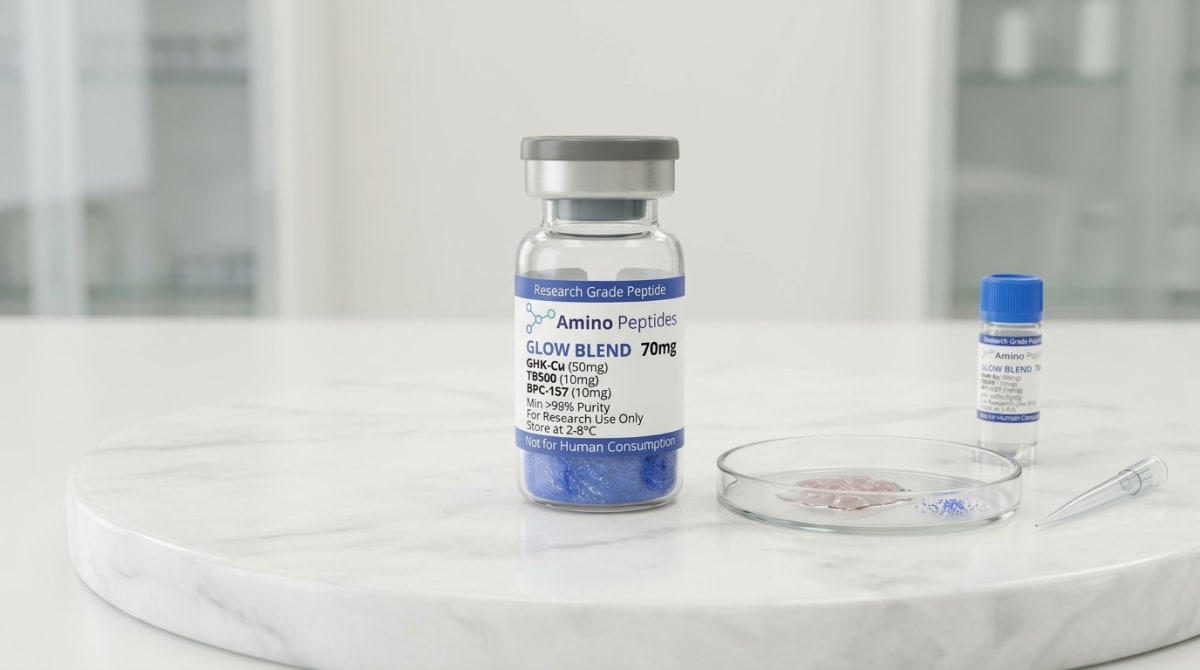
What You Need to Know About GLOW BLEND
- Researchers search for this topic to understand how to stabilise light output across large multi-well plates without relying on rapid fluid dispensing hardware.
- The underlying mechanism relies on slowing down the enzymatic reaction or clearing inhibitory byproducts so the enzyme emits a steady glow rather than a quick flash.
- Published studies observe that adding coenzyme A prevents the build-up of dehydroluciferyl-adenylate, extending the signal half-life from seconds to several hours.
- Laboratory models demonstrate that temperature shifts and well-to-well cross-talk must be tightly controlled to characterise kinetic stability accurately across 96-well or 384-well plates.
How Light Signals Reveal Cellular Energy and Tissue Repair
Bioluminescent glow kinetics measure how cells produce steady light signals when specific genes switch on, directly linking enzymatic activity to cellular energy and structural recovery. By tracking this light output in tiny microplate wells, scientists can watch tissue remodelling pathways activate in real time without destroying the sample.
The characterisation of bioluminescent glow kinetics in microplate-based luciferase reporter assays gives researchers a clear, measurable way to watch cellular energy and recovery mechanisms at work. At the centre of this method is an enzyme called luciferase, famously found in fireflies and glowing sea creatures. When researchers attach the genetic code for luciferase to a specific cellular target, the cell produces the enzyme whenever that target pathway switches on. Add a chemical fuel called luciferin alongside adenosine triphosphate—the main cellular energy molecule—and the enzyme produces visible light. Because this chemical reaction requires adenosine triphosphate to fire, the brightness and duration of the light directly reflect how much cellular energy is available inside the microplate well.
In older setups, adding the fuel caused a bright, one-second flash that faded almost immediately. Modern glow-type reagents slow that reaction down by gently regulating how fast the enzyme processes its fuel. Instead of a quick spark, the microplate wells emit a steady, measurable glow that lasts for hours. This steady light output matters enormously when investigating peptides linked to skin elasticity and structural recovery. For example, if a research team wants to see how a specific amino acid sequence switches on collagen genes or encourages blood vessel formation, they can measure the steady glow across ninety-six or three hundred and eighty-four tiny wells at the exact same time.
The brightness tells the scientist exactly how strongly the cells are responding to a given concentration of a test compound. Because there is no external light bulb shining into the dish, there is almost zero background glare to muddy the numbers. Published studies rely on this clean signal-to-noise ratio to map out how different study amounts influence fibroblast activity, antioxidant defence, and cellular energy production. By translating silent genetic switches into a steady beam of light, microplate reporter assays turn hidden microscopic repair processes into clear, reliable numbers that any laboratory reader can capture and compare.
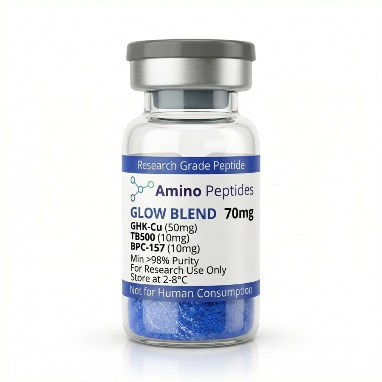
From £65
Supplied as a sealed freeze-dried vial (stable 12–24 months at 2°C–8°C in a standard refrigerator).
Shop GLOW BLEND →Tracking Flash Versus Steady Glow Across Study Timelines
Understanding the clock is everything when measuring light inside a multi-well plate. When scientists run a reporter assay, they are watching two separate clocks tick at the same time: the immediate chemical kinetics of the glowing enzyme, and the slower biological timeline of the cells responding to a test peptide. Getting reliable numbers means matching the right reading window to the right biological event.
In the first few seconds after luciferin hits the enzyme, the reaction spikes. In a flash-kinetic setup, peak light output happens within two to five seconds and drops steeply as reaction by-products like oxyluciferin build up and slow the enzyme down. To make large microplate experiments practical, modern assay buffers include coenzyme A and specialised stabilisers that clear away those bottlenecks. This chemical adjustment flattens the spike into a stable plateau—the classic glow phase—which typically holds steady from ten minutes post-mix out to two or three hours. During this window, the signal decays so slowly that a plate reader can scan hundreds of wells from top to bottom without the last well reading artificially lower than the first.
That chemical stability allows researchers to track the much longer timelines of cellular signalling and structural remodelling. Early signalling events happen fast. When a test peptide binds to a surface receptor in a laboratory model, internal messengers like cyclic adenosine monophosphate or calcium ions surge within five to thirty minutes. Secreted luciferase setups can capture these rapid shifts by sampling tiny drops of liquid from the well every fifteen minutes, leaving the living cells undisturbed at the bottom of the plate.
Later structural changes take much longer to show up on the reader. If an experiment is tracking how a peptide influences collagen synthesis, elastin cross-linking, or actin cytoskeleton reorganisation, the cells need time to transcribe DNA and build new proteins. In published studies, researchers typically incubate the cells with the test protocol ratio for twelve, twenty-four, or forty-eight hours before measuring the glow. Early six-hour readouts usually show the initial genetic switches flipping on, while the twenty-four to seventy-two-hour marks reveal sustained protein building and extracellular matrix assembly. Mapping both the early flash of signalling and the late plateau of structural assembly gives investigators a complete timeline of how cellular recovery unfolds from the first minute to the third day.
Comparison of Bioluminescent Reporter Assay Formats in Laboratory Models
| Assay Format | Primary Enzyme and Substrate | Signal Half-Life | Relative Photon Intensity | Hardware Requirement |
|---|---|---|---|---|
| Flash Luciferase Assay | Firefly luciferase with standard D-luciferin and ATP | Less than 1 minute | Very High (rapid peak then decay) | Luminometer with built-in automated fluid dispensers |
| Glow Firefly Luciferase Assay | Firefly luciferase with D-luciferin, ATP, and Coenzyme A | 2 to 5 hours | Moderate to High (steady plateau) | Standard microplate reader (batch processing) |
| NanoLuc Glow Assay | Engineered deep-sea shrimp luciferase with furimazine | Greater than 2 hours | Extremely High (bright and sustained) | Standard microplate reader with attenuation filters |
| Renilla Glow Assay | Sea pansy luciferase with protected coelenterazine analogues | 1 to 2 hours | Moderate (requires enzymatic deprotection) | Standard microplate reader |
| Dual-Reporter Glow System | Firefly and Renilla or NanoLuc enzymes in sequential buffers | 1 to 2 hours per phase | High (quench-and-read workflow) | Microplate reader with dual-wavelength or sequential reading |
Pairing Reporter Assays With Complementary Peptide Combinations
Bioluminescent reporter assays become even more informative when researchers test complementary peptide combinations rather than single sequences in isolation. In living tissue, structural recovery rarely relies on a single pathway; instead, cells coordinate actin movement, new blood vessel growth, and collagen deposition all at once. Microplate glow assays allow investigators to test how paired peptides interact across these distinct pathways by running side-by-side wells or dual-colour luciferase setups.
A popular area of tissue repair research involves pairing synthetic fragments of thymosin beta-4 with gastric pentadecapeptide sequences and copper-binding tripeptides. Each of these molecules targets a different stage of cellular upkeep. Thymosin beta-4 fragments, commonly studied as TB-500, bind to actin monomers to help cells migrate across gaps in a culture dish. Meanwhile, BPC-157 sequences are observed in preclinical models to influence growth factor receptor expression and nitric oxide signalling, while the copper tripeptide GHK-Cu supplies bioavailable copper ions that act as cofactors for lysyl oxidase, an enzyme that cross-links collagen and elastin fibres.
When scientists evaluate a bundled combination such as glow tb500 10mg in a multi-well reporter system, they can use dual-luciferase protocols to watch two genetic switches at the exact same time. For instance, firefly luciferase (which glows greenish-yellow) can be wired to a promoter for vascular endothelial growth factor, while a second enzyme from sea pansies, Renilla luciferase (which glows blue), is wired to a collagen type I promoter or used as an internal baseline control. By filtering the two light colours in the same microplate well, the assay shows whether combining the peptides produces a stronger, more sustained transcriptional response than testing each sequence alone.
Published studies using these multiplexed setups often observe that pairing an actin-regulating sequence with a copper-tripeptide complex smooths out the cellular response curve. While the migration-focused peptide triggers an early surge in cytoskeletal gene activity within the first six to twelve hours, the copper peptide supports a longer, steadier rise in matrix-building genes over forty-eight hours. Testing these combinations in high-density microplates lets researchers fine-tune protocol ratios and confirm that the paired sequences complement each other without competing for cellular energy reserves.
Reconstitution calculator
Choose a vial mass and a bacteriostatic reconstitution solution volume. The figure is the resulting concentration.
Concentration: 2.50 mg/mL
Per 0.01mL (10µL): 25.0 mcg
Open the full calculatorBacteriostatic reconstitution solutionWhy Reagent Purity and Cold Storage Protect Your Assay Data
Because bioluminescent glow assays are sensitive enough to detect femtomole amounts of an enzyme, the quality of the peptides and reagents entering the microplate well determines whether the final numbers can be trusted. Even tiny amounts of leftover synthesis chemicals can choke the luciferase enzyme or stress the cells, artificially dimming the light output and ruining an otherwise well-planned experiment.
The most common hidden culprit in low-grade peptide vials is trifluoroacetic acid, or TFA. During solid-phase synthesis, chemists use TFA to cleave the finished peptide chain from its resin support. If a supplier skips thorough salt-exchange and purification steps, residual TFA stays trapped in the freeze-dried powder. When that impure powder is dissolved and added to a microplate well, the acid shifts the local pH and damages cell membranes. Worse still, firefly luciferase is notoriously sensitive to pH drops and halide or acid anions; even trace TFA contamination can shift the colour of the emitted light toward red and slash the brightness of the glow kinetic curve. Sourcing peptides verified at ninety-nine percent or higher purity via High-Performance Liquid Chromatography (HPLC), alongside Mass Spectrometry confirmation and proper TFA removal, protects the assay from these chemical blind spots.
Storage and handling play an equally important role in keeping both the peptides and the reporter substrates intact. Freeze-dried peptide vials should be stored in a dedicated refrigerator between two degrees Celsius and eight degrees Celsius, or frozen at minus twenty degrees Celsius for long-term holding, away from bright light and moisture. Once a researcher prepares the vial using a clean bacteriostatic reconstitution solution, the liquid must stay chilled at two degrees Celsius to eight degrees Celsius and be handled gently—never shaken hard, as foaming can denature delicate amino acid chains.
Before the plate ever slides into the luminometer, the assay plates and reconstituted reagents should be brought to a steady room temperature, typically around twenty-two degrees Celsius, for thirty minutes. Luciferase reaction speed changes by nearly ten percent for every single degree of temperature shift. Combining ninety-nine percent pure, TFA-free peptides with strict cold-chain storage and even room-temperature equilibration ensures that every photon of light captured by the microplate reader reflects true biological signalling rather than vial contamination.
Frequently Asked Questions
Why do researchers look up the characterisation of bioluminescent glow kinetics in microplate-based luciferase reporter assays?
Researchers search for this process to measure cellular signalling and gene activity accurately inside laboratory models. By tracking how steady the light output remains over time, scientists can optimise their experimental setups. This ensures that every well in the microplate gives a reliable, comparable reading during high-throughput screening.
What is the typical timeline for observing glow kinetics in these laboratory assays?
A standard observation timeline runs from ten minutes to several hours after mixing the assay reagents. Unlike quick flash reactions that fade in seconds, glow kinetics provide a slow, steady light signal. Published studies show this extended window lets researchers analyse multiple microplates without losing signal strength.
Can you combine or stack multiple luciferase reporters in a single microplate well?
Yes, researchers frequently stack two different luciferase enzymes, such as firefly and Renilla, within the same microplate well. This dual-reporter method lets scientists track two separate biological events at once. Each enzyme uses a distinct chemical substrate and protocol ratio, allowing clear, separate light readings from one sample.
How do reagent purity and fridge storage affect luciferase assay performance?
High reagent purity and proper cold storage are vital for keeping the light signal bright and predictable. Storing reconstituted substrates in a standard fridge for too long causes chemical breakdown and weak luminescence. For best results, laboratory protocols recommend keeping aliquots frozen and avoiding repeated freeze-thaw cycles.
Bibliography
- Thorne N, Inglese J, Auld DS. Illuminating insights into firefly luciferase and other bioluminescent reporters used in chemical biology. Chemistry & Biology. 2010;17(6):646-657. View published research
- Hall MP, Unch J, Binkowski BF, et al. Engineered luciferase reporter from a deep sea shrimp utilizing a novel imidazopyrazinone substrate. ACS Chemical Biology. 2012;7(11):1848-1857. View published research
- Fraga H. Firefly luminescence: a historical perspective and recent developments. Photochemical & Photobiological Sciences. 2008;7(2):146-158. View published research
- Fan F, Wood KV. Bioluminescent assays for high-throughput screening. Assay and compound Development Technologies. 2007;5(1):127-136. View published research
⚠️ Research Use Only Disclaimer: All peptides and compounds are sold strictly for in-vitro laboratory research purposes only. Not intended for human or veterinary use, not a dietary supplement, and not approved to diagnose, treat, cure, or prevent any disease or condition. For use by qualified researchers in suitably equipped laboratory environments only. Amino Peptides Ltd operates in full compliance with UK MHRA regulations and applicable EU research chemical guidelines.