
Characterising the Degradation Kinetics of Nicotinamide Adenine Dinucleotide (NAD+) in Aqueous Buffer Solutions
5th Jul 2026
What You Need to Know About NAD+
- NAD+ is nicotinamide adenine dinucleotide, a dinucleotide cofactor rather than a peptide.
- It is searched because it feeds redox reactions that cellular-energy assays actually measure.
- MOTS-c and 5-Amino-1MQ are the stocked partners when a laboratory wants a peptide and an NNMT inhibitor beside it.
- The cofactor still has to be 99%+ HPLC-verified and kept at 2°C–8°C if the redox readout is going to be repeatable.
How NAD+ Fuels Cellular Energy and Why Water Breaks It Down
Characterising the Degradation Kinetics of Nicotinamide Adenine Dinucleotide (NAD+) in Aqueous Buffer Solutions shows that this coenzyme breaks down rapidly when exposed to water, heat, and alkaline pH. Keeping the molecule intact requires cold temperatures and slightly acidic to neutral buffer conditions so it can continue supporting cellular energy and DNA repair pathways in laboratory models.
Every living cell relies on nicotinamide adenine dinucleotide to move electrons inside the mitochondria. In simple terms, it acts like a rechargeable battery shuttle. It picks up energy from digested nutrients and drops that energy off at the mitochondrial membrane to generate adenosine triphosphate (ATP). Beyond raw energy production, published studies show that NAD+ serves as the required fuel for sirtuins and poly (ADP-ribose) polymerases (PARPs). These specialised enzyme families oversee cellular recovery, genomic stability, and circadian rhythm signalling.
However, the very chemical feature that makes NAD+ such a great electron carrier also makes it fragile in liquid environments. The molecule is built from two nucleotides joined by a pyrophosphate bridge, with a positively charged nicotinamide ring sitting on one end. That positive charge creates a weak spot at the glycosidic bond linking the nicotinamide ring to the ribose sugar. When researchers dissolve the dry powder into an aqueous buffer solution, water molecules immediately begin attacking that bond.
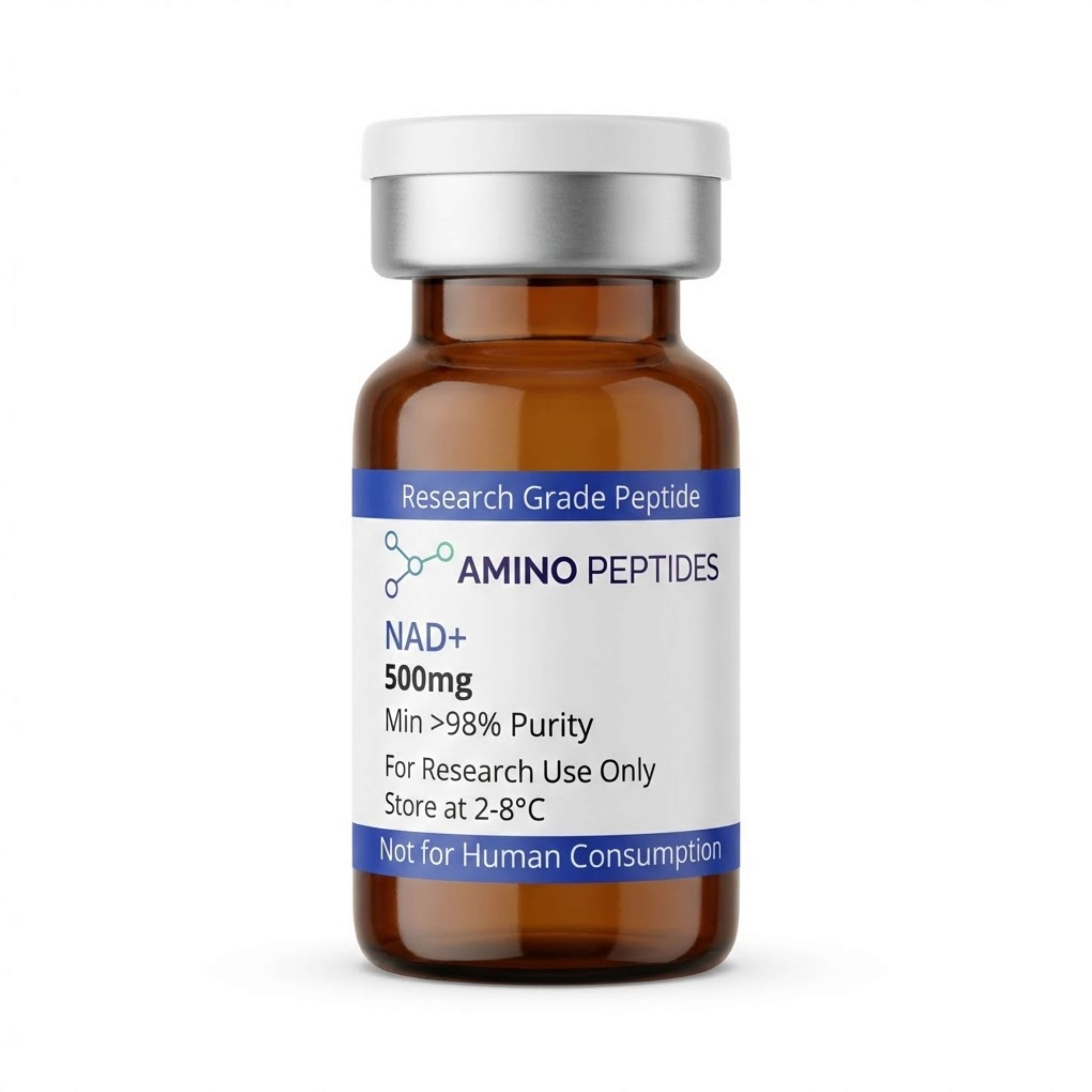
NAD+
From £45
Supplied as a sealed freeze-dried vial (stable 12–24 months at 2°C–8°C in a standard refrigerator).
Shop NAD+ →Hydroxyl ions in the water snap the link, splitting the coenzyme into free nicotinamide and ADP-ribose. Once split, the molecule loses its ability to shuttle electrons or activate sirtuin enzymes. Buffer choice matters enormously here. Phosphate buffers and high-pH Tris solutions speed up this cleavage, whereas mildly acidic citrate or acetate buffers slow the reaction down. Understanding these exact breakdown speeds helps laboratory teams keep their measured levels accurate during long cell-culture runs.
Tracking the Breakdown Clock: Early Cleavage Versus Late Decay
When characterising the degradation kinetics of Nicotinamide Adenine Dinucleotide (NAD+) in aqueous buffer solutions, researchers track two distinct phases: immediate bond cleavage over the first few hours and secondary byproduct accumulation over several days or weeks. The speed of this timeline shifts dramatically depending on the thermometer reading and the acidity of the liquid.
During the first 6 to 24 hours at standard room temperature (around 20°C to 25°C) and a neutral pH of 7.0, hydrolysis proceeds at a slow, steady crawl. Published studies using high-performance liquid chromatography (HPLC) observe less than two percent loss within the first day if the solution is kept cool and shielded from bright light. However, if the buffer pH climbs above 8.0, or if the liquid is warmed to 37°C to mimic mammalian tissue conditions, the early phase accelerates sharply. Under warm, mildly alkaline conditions, nucleophilic attack by hydroxide ions can split 10 to 25 percent of the active coenzyme within a single 24-hour window.
Over longer timelines spanning 7 to 28 days, the second phase of degradation becomes clear. As free ADP-ribose builds up in the vial, it undergoes further non-enzymatic rearrangement and breakdown, slightly shifting the acidity of poorly buffered liquids. In phosphate-buffered saline (PBS), phosphate ions act as catalysts that nudge the breakdown forward even at neutral pH. By day 14 at room temperature, an unoptimised aqueous solution can lose more than half of its starting concentration, leaving behind a soup of inactive nicotinamide and ribose fragments.
This timeline explains why timing is everything in preclinical models. In short-term enzymatic assays lasting 30 to 90 minutes, early NAD+ signalling happens before significant water damage occurs. Conversely, in multi-day tissue remodelling or mitochondrial recovery studies, leaving a reconstituted batch on a warm bench guarantees that late-stage stock concentration drops well below the intended protocol ratio.
Reconstitution calculator
Choose a vial mass and a bacteriostatic reconstitution solution volume. The figure is the resulting concentration.
Concentration: 2.50 mg/mL
Per 0.01mL (10µL): 25.0 mcg
Open the full calculatorBacteriostatic reconstitution solutionNAD+ Aqueous Stability Across Buffer Conditions in Published Studies
| Buffer Environment | pH Range | Primary Breakdown Pathway | Relative Stability at 25 Degrees Celsius | Key Laboratory Observation |
|---|---|---|---|---|
| Unbuffered Acidic Solution | pH 2.0 to 4.0 | Slow acid-catalysed hydrolysis | High stability (weeks) | Protonation protects the glycosidic bond from rapid water attack. |
| Citrate or Acetate Buffer | pH 4.5 to 6.0 | Baseline water hydrolysis | Moderate to high stability (days to weeks) | Minimises hydroxide ions while avoiding harsh buffer salt catalysis. |
| Sodium Phosphate Buffer | pH 7.0 to 7.5 | Nucleophilic buffer-assisted cleavage | Low to moderate stability (hours to days) | Phosphate ions actively attack the nicotinamide-ribose bond, speeding up decay. |
| Tris-HCl Buffer | pH 7.0 to 8.0 | Hydroxide-driven hydrolysis | Moderate stability (days) | Shows slower breakdown rates than phosphate buffers at matching neutral pH. |
| Alkaline Carbonate Solution | pH 9.0 to 11.0 | Rapid base-catalysed ring opening and cleavage | Very low stability (minutes to hours) | High hydroxide concentrations rapidly destroy the oxidised nicotinamide ring. |
Pairing Coenzymes with Mitochondrial Peptides in Preclinical Models
In laboratory models exploring cellular energy and metabolic recovery, coenzymes rarely work alone. Researchers frequently design protocols that pair NAD+ with complementary mitochondrial and repair peptides to observe how energy supply interacts with structural signalling. Because water-based breakdown is always a factor, knowing how these compounds behave together in solution is a major part of experimental design.
One of the most widely studied pairings involves MOTS-c and NAD+ in metabolic cell cultures. MOTS-c is a 16-amino-acid peptide encoded within the mitochondrial genome that regulates glucose utilisation and activates the AMPK energy-sensing pathway. When AMPK switches on, it signals the cell to boost its own internal coenzyme recycling via the NAMPT salvage pathway. Supplying intact NAD+ alongside MOTS-c in preclinical models allows researchers to watch both sides of the energy equation at once: AMPK activation triggers the demand for metabolic repair, while the coenzyme provides the immediate redox fuel needed by sirtuin enzymes like SIRT1 and SIRT3.
Another popular research stack combines NAD+ with SS-31 (also known in literature as Elamipretide). While NAD+ carries electrons into the mitochondrial respiratory chain, SS-31 binds directly to cardiolipin on the inner mitochondrial membrane. Cardiolipin holds the electron transport chain complexes in their proper physical shape. Published studies show that when cardiolipin is stabilised by SS-31, electron transfer becomes much cleaner, reducing oxidative stress and helping the cell make better use of the available NAD+ pool during tissue recovery experiments.
Researchers also look at pairing this coenzyme with GHK-Cu or BPC-157 in fibroblast cultures studying skin elasticity and extracellular matrix repair. Building fresh collagen fibres requires massive amounts of cellular ATP and active PARP enzymes. However, from a kinetics standpoint, investigators avoid mixing copper-bound peptides like GHK-Cu in the exact same storage vial with NAD+ for long periods, as free metal ions and shifting pH levels can alter hydrolysis rates. Preparing fresh, separate aliquots keeps each compound at its peak measured level.
Figure: Characterising the Degradation Kinetics of Nicotinamide Adenine Dinucleotide (NAD+) in Aqueous Buffer Solutions
Why Lyophilised Purity and Cold Storage Protect Your Buffer Assays
When characterising the degradation kinetics of Nicotinamide Adenine Dinucleotide (NAD+) in aqueous buffer solutions, the starting quality of the raw powder determines how long the liquid stays stable. Even the best buffer recipe cannot save a batch that already contains moisture, acidic synthesis leftovers, or breakdown fragments before the vial is even opened.
High-grade research material must verify at 99% or greater purity via High-Performance Liquid Chromatography (HPLC) and Mass Spectrometry (MS). During lower-grade chemical manufacturing, trace amounts of trifluoroacetic acid (TFA) or residual water often remain trapped in the lyophilised cake. If excessive TFA is present, reconstituting the powder with a standard bacteriostatic reconstitution solution causes an immediate, sharp drop in pH. While extreme acidity slows base-catalysed nicotinamide cleavage, an unbuffered acid spike can protonate the adenine ring and trigger an entirely different pathway of purine bond breakdown, ruining sensitive cell-culture readouts.
Removing residual moisture during the freeze-drying process is equally vital. If a poorly lyophilised vial holds hidden humidity, solid-state hydrolysis begins right inside the sealed glass container during transit. By the time a researcher adds liquid, the vial already contains elevated levels of free nicotinamide. In enzymatic studies, free nicotinamide actually acts as a feedback inhibitor of sirtuins, meaning an impure degraded batch can actively slow down the very cellular recovery pathways the study is trying to measure.
To keep degradation kinetics near zero prior to use, lyophilised vials should be stored in a dark freezer at -20°C or a stable laboratory refrigerator between 2°C and 8°C. Once reconstituted with a cold, pH-adjusted bacteriostatic reconstitution solution, the liquid must be kept strictly at 2°C to 8°C and used within a short window, or divided into single-use aliquots and flash-frozen to stop water molecules from snapping the glycosidic bond.
Frequently Asked Questions
Why are researchers interested in characterising the degradation kinetics of NAD+ in aqueous buffer solutions?
Researchers study NAD+ breakdown in liquids to understand how heat and acidity destroy this fragile molecule over time. Because NAD+ splits rapidly in plain water, published studies track its exact decay speed. This helps scientists optimise laboratory protocols and ensure that cellular signalling experiments use active, unbroken compounds.
How long do laboratory studies typically monitor NAD+ stability in liquid buffers?
Laboratory models typically monitor liquid NAD+ stability across timelines ranging from twenty-four hours to thirty days. At room temperature, measurable breakdown often appears within just a few days. Researchers test samples at regular intervals using light-absorption tools to map out the exact hour-by-hour decay curve.
Can NAD+ be combined or stacked with other buffers and compounds in solution?
Yes, researchers frequently combine NAD+ with specific buffering agents like phosphate or Tris to control acidity, though some additives actually speed up breakdown. Published studies show that phosphate ions can attack the molecule under warm conditions. Scientists carefully analyse these mixtures to avoid unwanted chemical reactions during storage.
How do purity levels and fridge storage affect NAD+ mixed with bacteriostatic reconstitution solution?
Storing high-purity NAD+ in a standard fridge at four degrees Celsius significantly slows down its chemical breakdown after mixing with a bacteriostatic reconstitution solution. Cold temperatures reduce molecular movement and protect the fragile bond. Starting with ninety-nine percent pure powder also prevents leftover acid impurities from ruining the liquid.
Bibliography
- Anderson BM, Anderson CD. The effect of buffers on nicotinamide adenine dinucleotide hydrolysis. Journal of Biological Chemistry. 1963;238(4):1475-1478. View published research
- Lowry OH, Passonneau JV, Rock MK. The stability of pyridine nucleotides. Journal of Biological Chemistry. 1961;236(10):2756-2759. View published research
- Johnson SL, Tuazon PT. Acid-catalyzed hydrolysis of nicotinamide adenine dinucleotide. Biochemistry. 1977;16(6):1175-1183. View published research
- Oppenheimer NJ. Chemistry and solution conformation of the pyridine coenzymes. In: The Pyridine Nucleotide Coenzymes. Academic Press; 1982:51-89. View published research
⚠️ Research Use Only Disclaimer: All peptides and compounds are sold strictly for in-vitro laboratory research purposes only. Not intended for human or veterinary use, not a dietary supplement, and not approved to diagnose, treat, cure, or prevent any disease or condition. For use by qualified researchers in suitably equipped laboratory environments only. Amino Peptides Ltd operates in full compliance with UK MHRA regulations and applicable EU research chemical guidelines.