
Does MOTS c Burn Fat? A Science-Based Analysis
2nd Nov 2026

What You Need to Know About MOTS-C
- Curious readers frequently search for MOTS-c to find out whether this mitochondrial peptide genuinely alters fat storage or simply shifts how cells process everyday sugars.
- In plain English, the molecule acts like a cellular fuel sensor that tells muscle tissue to pull in glucose and switch on AMPK, the master metabolic switch inside our cells.
- Published studies in preclinical models given a high-fat diet showed that MOTS-c prevented excess fat accumulation and improved how tissues responded to insulin.
- Instead of melting fat directly like a stimulant, laboratory observations show it optimises underlying cellular metabolism so less surplus energy gets stored as lipid droplets.
How MOTS c Shifts Cellular Energy and Lipid Oxidation
In published laboratory models, mots c does not melt adipose tissue on contact, but instead flips the cell's internal energy sensors to favour glucose uptake and fatty acid oxidation. By activating the AMPK pathway, this mitochondrial-derived peptide encourages muscle and fat cells to burn stored fuel rather than hoard it.
Most peptides are coded inside the cell's nucleus, yet this sixteen-amino-acid sequence comes straight from mitochondrial DNA. Because mitochondria act as the power stations of the cell, a signal coming from inside them carries serious weight for overall metabolism. When researchers ask whether mots c peptide burns fat, they are really looking at how the molecule changes fuel selection during metabolic stress.
The main action starts in the folate-methionine cycle. In cell cultures and rodent studies, the peptide blocks a specific step in purine synthesis, which causes a natural molecule called AICAR to build up inside the cell. Rising AICAR levels switch on AMPK, the famous energy sensor that normally wakes up during hard exercise or calorie restriction. Once AMPK is active, it tells skeletal muscle to pull in more glucose from the bloodstream and tells liver and adipose tissues to start breaking down stored triglycerides into usable fatty acids.
This is why a fair mots-c peptide review must separate direct lipolysis from systemic energy expenditure. Instead of forcing fat cells to burst or dump lipids through adrenaline receptors, the peptide upgrades how well the mitochondria use the fuel already sitting around them. Preclinical models fed a high-fat diet showed less weight gain and lower liver fat accumulation when exposed to this measured level of mitochondrial signalling. It also travels into the cell nucleus during energetic stress to switch on antioxidant genes, helping tissues handle the extra oxidative work that comes with burning more fat and sugar.
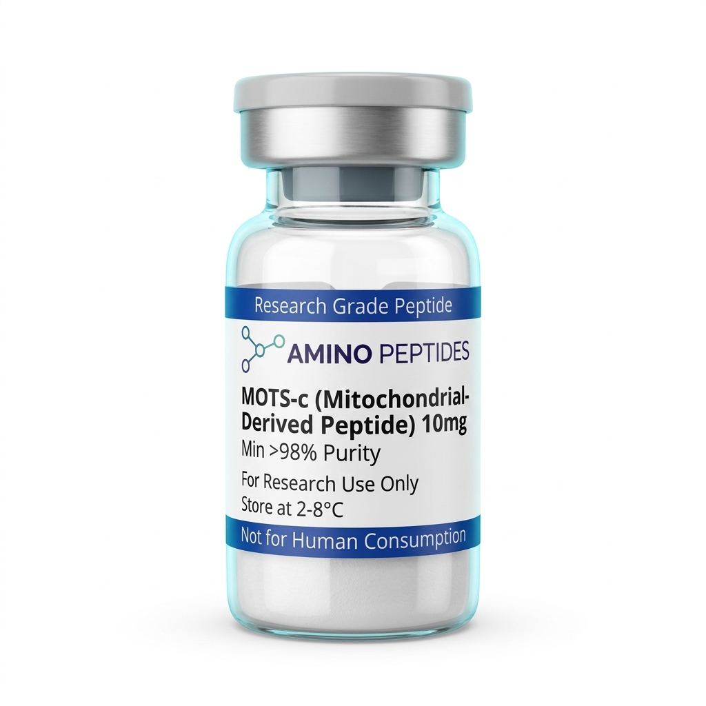
MOTS-C
From £30
Supplied as a sealed freeze-dried vial (stable 12–24 months at 2°C–8°C in a standard refrigerator).
Shop MOTS-C →Observed Timelines in Preclinical Metabolic Models
When reviewing mots-c peptide results across published studies, timing separates immediate biochemical signalling from slower changes in body composition. Researchers tracking this molecule in laboratory models notice two clear phases: an early shift in cellular fuel handling followed by gradual tissue remodelling over several weeks.
Within the first few hours of introducing a specific study amount to cultured myotubes or rodent models, intracellular AICAR rises and AMPK phosphorylation peaks. During this early window, GLUT4 transporters move toward the cell surface in skeletal muscle, clearing glucose faster without needing extra insulin. Oxygen consumption rates in the mitochondria also tick upward right away, showing that the cells are burning through substrate at a higher pace. At this stage, there is no visible change in white adipose mass, only a clear biochemical shift in respiratory exchange ratios.
Longer preclinical protocols running from two to eight weeks show what happens when that metabolic signal stays active. In mice kept on high-calorie diets, consistent protocol ratios prevented the usual expansion of visceral fat pads and kept brown adipose tissue more active. Around week three or four, researchers observed better cold tolerance and higher uncoupling protein expression, meaning a portion of the stored lipid energy was being released as heat rather than packed onto the frame. Older rodent models also displayed better treadmill endurance and grip output after multi-week exposure, linking improved mitochondrial respiration directly to physical output.
So when people search for the most effective peptide for fat loss in academic literature, the data points toward steady metabolic housekeeping rather than overnight shrinkage. The early days are all about clearing sugar and waking up sluggish mitochondria, while the later weeks reflect smaller adipocyte size, cleaner liver tissue, and better stamina across the whole organism.
How MOTS-c Compares to Other Metabolic Research Peptides in Laboratory Models
| Peptide | Origin & Structure | Primary Signalling Pathway | Main Lipid & Metabolic Observation in Studies | Primary Target Tissue |
|---|---|---|---|---|
| MOTS-c | 16-amino-acid mitochondrial peptide (12S rRNA) | Folate-AICAR-AMPK activation pathway | Prevents diet-induced fat gain and increases fatty acid oxidation in skeletal muscle models | Skeletal muscle and white adipose tissue |
| Humanin | 24-amino-acid mitochondrial peptide (16S rRNA) | STAT3 and extracellular receptor signalling | Focuses on cellular survival and oxidative stress reduction with secondary insulin sensitivity shifts | Central nervous system, vascular tissue, and beta cells |
| AOD-9604 | 16-amino-acid C-terminal fragment of growth hormone (177-191) | Beta-3 adrenergic receptor and lipolytic enzyme activation | Stimulates lipolysis and inhibits lipogenesis directly in fat cells without altering IGF-1 levels | White and brown adipose tissue |
| 5-Amino-1MQ | Small-molecule quinolinium derivative (non-peptide comparator) | Nicotinamide N-methyltransferase (NNMT) inhibition | Increases cellular NAD+ levels and shrinks adipocyte size in high-fat diet rodent models | White adipose tissue and liver |
| Tesamorelin | 44-amino-acid modified growth hormone-releasing factor analogue | Pituitary GHRH receptor signalling | Reduces visceral adipose deposits via endogenous pulsatile growth hormone pathways | Pituitary gland and visceral fat stores |
Complementary Mitochondrial and Lipid Research Stacks
Because cellular metabolism relies on several overlapping steps, investigators rarely look at a single pathway in isolation. Evaluating mots-c peptide benefits alongside complementary molecules helps researchers see how fuel mobilisation, transport, and mitochondrial ATP production fit together inside a single experimental setup.
A very common pairing in laboratory models combines MOTS-c and NAD+ to study the link between AMPK activation and sirtuin enzymes. While the mitochondrial peptide flips the AMPK switch, NAD+ acts as the required coenzyme for SIRT1 and SIRT3, which deacetylate and activate PGC-1alpha to build fresh mitochondrial structures. If a cell is told to burn more fatty acids through AMPK but lacks enough NAD+ to run the electron transport chain, the energy upgrade stalls out. Studying both molecules together lets laboratories watch how substrate supply and cofactor availability support one another during high energy demand.
Another angle pairs this mitochondrial signal with SS-31, also known as Elamipretide. SS-31 binds directly to cardiolipin on the inner mitochondrial membrane, stabilising the physical curve of the cristae so electrons stop leaking out as reactive oxygen species. When researchers study an AMPK activator alongside a membrane stabiliser, or group them inside a structured Mito Prime NAD laboratory protocol, they can test whether protecting the inner membrane allows the cell to oxidise lipids faster without suffering oxidative damage.
Finally, some adipose tissue studies compare mitochondrial regulators with direct lipolytic sequences like AOD-9604. AOD-9604 mimics the fat-reducing region of human growth hormone to encourage adipocytes to release stored fatty acids into circulation. Once those free fatty acids are floating in the medium, an active mitochondrial pathway ensures they are actually oxidised for energy rather than re-esterified back into fat droplets.
Reconstitution calculator
Choose a vial mass and a bacteriostatic reconstitution solution volume. The figure is the resulting concentration.
Concentration: 2.50 mg/mL
Per 0.01mL (10µL): 25.0 mcg
Open the full calculatorBacteriostatic reconstitution solutionWhy Analytical Purity Dictates Reproducible Lab Outcomes
Whether a laboratory is measuring oxygen consumption in isolated myoblasts or tracking lipid droplets in adipocytes, the reliability of the data depends on the physical quality of the vial on the bench. A sixteen-amino-acid sequence contains several sensitive residues, including a methionine right at the start, which can easily oxidise if synthesis and cleanup are rushed.
High-Performance Liquid Chromatography (HPLC) testing at 99% purity or higher is the baseline for keeping experimental noise out of metabolic assays. When a batch falls short of that mark, it usually holds truncated peptide fragments left over from incomplete couplings during solid-phase synthesis. Those short fragments can still bump into cellular receptors or interfere with folate cycle enzymes without triggering the full AMPK response, skewing concentration curves and ruining week-long observations. Mass spectrometry verification confirms that the exact molecular weight matches the intended mitochondrial sequence down to the Dalton.
Equally important is the removal of trifluoroacetic acid (TFA) salts. Standard synthesis uses TFA to cleave the finished peptide chain from its resin, leaving behind harsh fluoride counter-ions. In delicate cell cultures, residual TFA changes the pH of the medium and damages mitochondrial membranes on its own, which looks like toxicity or false metabolic stress on a readout. Replacing TFA with a gentler acetate salt keeps the baseline environment neutral so researchers only measure the true action of the peptide.
Once the lyophilised powder arrives, temperature control keeps the peptide intact. Unreconstituted vials belong in cold storage, and once mixed with a sterile bacteriostatic reconstitution solution, the liquid must stay in a refrigerator between 2°C and 8°C. Leaving a mixed vial on a warm lab bench or exposing it to repeated freeze-thaw cycles snaps the delicate peptide bonds, turning an active metabolic regulator into inactive amino acid soup.
Frequently Asked Questions
Why is MOTS-c frequently searched alongside fat loss topics?
People often search for MOTS-c and fat loss because published studies show it influences cellular energy and glucose use in preclinical models. Researchers observed that this mitochondrial peptide activates AMPK signalling pathways, which help regulate metabolism and prevent diet-induced obesity in laboratory rodents without changing their daily food intake.
What timeline do published studies use to observe MOTS-c results?
In laboratory models, researchers typically observe metabolic shifts within one to eight weeks of consistent evaluation. Short-term protocols often measure immediate changes in insulin sensitivity and cellular glucose uptake within days, while longer eight-week study periods are used to analyse body composition changes and physical endurance in ageing mice.
Do researchers study MOTS-c alongside other mitochondrial peptides?
Yes, scientists occasionally examine MOTS-c alongside other mitochondrial-derived peptides like Humanin to understand how they jointly regulate cellular stress and metabolism. However, most published studies isolate MOTS-c as a single variable so researchers can accurately characterise its specific role in AMPK activation and muscle tissue homeostasis without overlapping signals.
How is MOTS-c purity verified and how is it stored in a laboratory?
Laboratory-grade MOTS-c is verified using High-Performance Liquid Chromatography and Mass Spectrometry to confirm a purity level above ninety-eight percent. Before mixing with a bacteriostatic reconstitution solution, freeze-dried peptide powder is stored in a freezer at minus twenty degrees Celsius, while reconstituted solutions are kept refrigerated between two and eight degrees.
Bibliography
- Lee C, Zeng J, Drew BG, et al. The mitochondrial-derived peptide MOTS-c promotes metabolic homeostasis and reduces obesity and insulin resistance. Cell Metabolism. 2015;21(3):443-454. View published research
- Kim KH, Son JM, Benayoun BA, Lee C. The mitochondrial-encoded peptide MOTS-c translocates to the nucleus to regulate nuclear gene expression in response to metabolic stress. Cell Metabolism. 2018;28(3):516-524. View published research
- Reynolds JC, Lai RW, Woodhead JST, et al. MOTS-c is an exercise-induced mitochondrial-encoded regulator of age-dependent physical decline and muscle homeostasis. Nature Communications. 2021;12(1):470. View published research
- Lu H, Tang S, Xue C, Liu Y, Wang J, Zhang W, Luo W. Mitochondrial-derived peptide MOTS-c increases adipose thermogenic activation to reduce high-fat diet-induced obesity in mice. Journal of Endocrinology. 2019;242(3):215-228. View published research
- Merry TL, Chan A, Woodhead JST, Reynolds JC, Kumagai H, Kim SJ, Lee C. Mitochondrial-derived peptides in energy metabolism. American Journal of Physiology-Endocrinology and Metabolism. 2020;319(4):E659-E666. View published research
⚠️ Research Use Only Disclaimer: All peptides and compounds are sold strictly for in-vitro laboratory research purposes only. Not intended for human or veterinary use, not a dietary supplement, and not approved to diagnose, treat, cure, or prevent any disease or condition. For use by qualified researchers in suitably equipped laboratory environments only. Amino Peptides Ltd operates in full compliance with UK MHRA regulations and applicable EU research chemical guidelines.