
GHK-Cu, BPC-157 and TB-500: Fibroblast Migration Synergy
27th Aug 2026

In cellular biology, complex chemical signals direct how cells maintain and repair structural tissues. In laboratory settings, researchers constantly analyse how different compounds influence the behaviour of isolated cells. A current area of scientific focus involves testing combinations of synthetic peptides to see if they produce a synergistic effect. Specifically, scientists are examining how a mixture of GHK-Cu, BPC-157, and TB-500 alters the activity of dermal fibroblasts in controlled cell cultures.
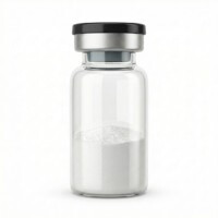
BPC-157
HPLC-verified lyophilised compound, UK-domestic supply. Manufactured under controlled conditions for qualified in-vitro laboratory research.
View Reagent Profile ›Dermal fibroblasts are the primary builder cells found in the structural layer of the skin. In a petri dish, these cells are responsible for producing the extracellular matrix, which is the biological scaffold made of collagen and elastin. When researchers introduce specific peptides to these isolated cells, they observe distinct changes in how the cells move, multiply, and express their genes.

Figure 1: Synergistic Effects of a GHK-Cu, BPC-157, and TB-500 Peptide Combination on Dermal Fibroblast Migration and Extracellular Matrix Gene Expression In Vitro
Key Takeaways
- Dermal fibroblasts change their physical shape when exposed to specific peptide combinations.
- TB-500 primarily influences the internal actin skeleton of the cell to facilitate movement.
- BPC-157 acts as a survival and signalling molecule during cellular stress assays.
- GHK-Cu directly impacts the gene expression required for collagen production.
- Combining these three peptides in vitro results in faster cell migration across a culture dish.
To understand the synergistic effect, researchers first look at the isolated role of each peptide. TB-500 is a synthetic fraction of Thymosin Beta-4. In cellular models, it binds to actin. Actin is a protein that forms the internal skeleton of the cell. By interacting with actin, TB-500 allows the cell to change its shape rapidly, which is a mechanical requirement for physical movement across a surface.
Before conducting these cellular assays, laboratory technicians must verify the exact molecular identity of the BPC-157 pentadecapeptide being used. They achieve this by cross-referencing the batch certificate of analysis with the official specification sheet to confirm purity and mass. Once verified, BPC-157 is introduced to the culture. This pentadecapeptide functions primarily as a signalling molecule. Laboratory data shows that it promotes cell survival under stress and accelerates the rate at which cells extend their leading edges to travel.
The third component, GHK-Cu, is a copper-binding tripeptide. While TB-500 and BPC-157 focus heavily on cellular movement and survival, GHK-Cu targets the manufacturing centre of the cell. In vitro studies demonstrate that GHK-Cu upregulates the specific genes responsible for producing collagen and elastin. It essentially instructs the cell to begin manufacturing the building blocks of the extracellular matrix.
When researchers combine these three peptides, they must first dissolve the lyophilised powders in a precise bacteriostatic reconstitution solution to ensure molecular stability. Once the combined solution is applied to a cell culture, scientists use a technique called a scratch assay to measure the results. They grow a solid layer of fibroblasts in a dish and drag a microscopic tool down the middle to create an empty gap. They then time exactly how long it takes the cells to cross the empty space.
In laboratory scratch assays, this coordinated cellular response demonstrates that peptide combinations can trigger multiple biological pathways simultaneously.
In the control groups, cells cross the gap at a standard, predictable rate. However, in the groups exposed to the peptide combination, the cells cross the gap significantly faster. The laboratory data suggests a clear division of labour among the molecules. TB-500 provides the mechanical flexibility for movement, BPC-157 provides the chemical signal to accelerate that movement, and GHK-Cu ensures that once the cells arrive in the empty space, they immediately begin expressing the genes necessary to build a new matrix.
Frequently Asked Questions: In-Vitro Fibroblast Dynamics
What is dermal fibroblast morphology?
In laboratory terms, dermal fibroblast morphology refers to the physical shape and structure of the cell. Under a microscope, a resting fibroblast typically looks flat and star-shaped. When activated by peptides, its morphology changes; it stretches out and becomes spindle-shaped, which allows it to move efficiently across the culture dish.
How does dermal fibroblast migration occur?
Dermal fibroblast migration occurs when the cell alters its internal actin skeleton to push itself forward. The cell extends a leading edge, grips the surface of the petri dish, and then pulls its trailing end forward. Researchers measure this exact process using scratch assays.
What acts as a fibroblast mitogen?
A fibroblast mitogen is any chemical signal that instructs the cell to divide and multiply. In controlled assays, specific growth factors and synthetic peptides can act as mitogens, increasing the total number of active cells available to participate in matrix construction.
What defines a migrating fibroblast?
A migrating fibroblast is a cell that has shifted from a stationary, protein-building state into an active, moving state. It stops focusing entirely on secreting collagen and instead redirects its energy towards physical movement, relying heavily on actin filaments to navigate its environment.
What role do dermal fibroblasts play in wound closure models?
When researchers study dermal fibroblasts in wound closure models in vitro, they observe these cells acting as the primary construction crew. The cells migrate into the artificially damaged area of the cell culture, multiply, and secrete the proteins required to build a new structural matrix.
Scientific Bibliography
- Pickart, L., et al. (2015). GHK peptide as a natural modulator of multiple cellular pathways in skin regeneration. BioMed Research International, 2015, 648108. View published research
- Philp, D., et al. (2004). Thymosin beta4 promotes angiogenesis, wound healing, and hair follicle development. Annals of the New York Academy of Sciences, 1028, 344-353. View published research
- Tkalcevic, V. I., et al. (2007). Enhancing effect of pentadecapeptide BPC 157 on wound healing and angiogenesis in rats. European Journal of Pharmacology, 577(1-3), 212-219. View published research
- Kang, E. A., et al. (2018). BPC157 as potential agent rescuing from cancer cachexia. Current Pharmaceutical Design, 24(18), 1947-1956. View published research
- Maquart, F. X., et al. (1988). Stimulation of collagen synthesis in fibroblast cultures by the tripeptide-copper complex glycyl-L-histidyl-L-lysine-Cu2+. FEBS Letters, 238(2), 343-346. View published research
- Sosne, G., et al. (2010). Thymosin beta4: structure, function, and biological properties. Annals of the New York Academy of Sciences, 1194, 200-210. View published research
- Hsieh, M. J., et al. (2017). Therapeutic potential of pro-angiogenic BPC157 is associated with VEGFR2 activation and up-regulation. Journal of Molecular Medicine, 95(3), 323-333. View published research
⚠️ Research Use Only Disclaimer: All peptides and compounds are sold strictly for in-vitro laboratory research purposes only. Not intended for human or veterinary use, not a dietary supplement, and not approved to diagnose, treat, cure, or prevent any disease or condition. For use by qualified researchers in suitably equipped laboratory environments only. Amino Peptides Ltd operates in full compliance with UK MHRA regulations and applicable EU research chemical guidelines.
Independent, batch-specific documentation for BPC-157 — reviewed prior to publication for in-vitro laboratory research and molecular stability reference.