
Hexarelin: The Most Potent GHS for Cardiovascular Research?
28th Sep 2026

What You Need to Know About Hexarelin
- Researchers frequently investigate Hexarelin to understand why it triggers a stronger growth hormone pulse than almost any other synthetic peptide in its class.
- It works through a unique dual pathway by binding to both the standard ghrelin receptor (GHS-R1a) and a specific cardiac scavenger receptor known as CD36.
- Published laboratory models observe that its interaction with CD36 supports heart muscle survival, preserves left ventricular output, and reduces fibrotic scarring independently of growth hormone release.
- Because it also raises cortisol and prolactin levels and causes rapid receptor desensitisation in animal assays, laboratory protocols must carefully time exposure windows.
How Hexarelin Drives Heart Tissue Recovery through Dual Receptor Binding
Hexarelin is a six-amino-acid synthetic peptide that triggers strong growth hormone release while simultaneously binding directly to specialised CD36 receptors on heart muscle cells. Because it acts through both pituitary signalling and local cardiac receptors, published studies rank it among the strongest growth hormone secretagogues for examining cardiovascular tissue recovery and cellular energy preservation.
Most compounds in the growth hormone secretagogue (GHS) family do one main job: they mimic the hunger hormone ghrelin and bind to the GHS-R1a receptor in the pituitary gland to prompt a pulse of growth hormone. Hexarelin does that with high intensity, often outperforming earlier peptides like GHRP-6 and GHRP-2 on a weight-for-weight basis in laboratory models. Yet what makes researchers ask whether it is the most potent GHS for cardiovascular work is a second, completely independent mechanism.
On the surface of cardiomyocytes—the working muscle cells of the heart—and endothelial cells lining blood vessels sits a scavenger receptor called CD36. When scientists test Hexarelin research peptide in isolated heart preparations, even in models bred without a pituitary gland, the peptide still protects heart muscle cells from low-oxygen stress. It does this by latching onto CD36 and keeping the cell's energy factories, the mitochondria, from swelling and breaking down during periods of oxygen deprivation followed by blood flow return.
In plain English, when heart tissue is starved of oxygen in a dish or an animal model, calcium floods into the cells and triggers self-destruct signals known as apoptosis. Published studies show that Hexarelin slows down that calcium overload and supports survival proteins like Bcl-2 while turning down cell-death markers like Bax and caspase-3. At the same time, its interaction with CD36 helps regulate how fatty acids are taken up by heart tissue, keeping cellular energy steady rather than chaotic. For researchers studying recovery, vascular elasticity, and cellular stamina under stress, this dual action—systemic growth hormone release plus direct local CD36 protection—sets Hexarelin apart from nearly every other secretagogue in the catalogue.
Because the third amino acid in its chain is a methylated tryptophan, the molecule resists enzymatic breakdown longer than natural ghrelin. That simple structural tweak gives laboratory models a steadier signalling window to observe both pituitary output and direct heart cell preservation without the rapid drop-off seen in unmodified sequences.
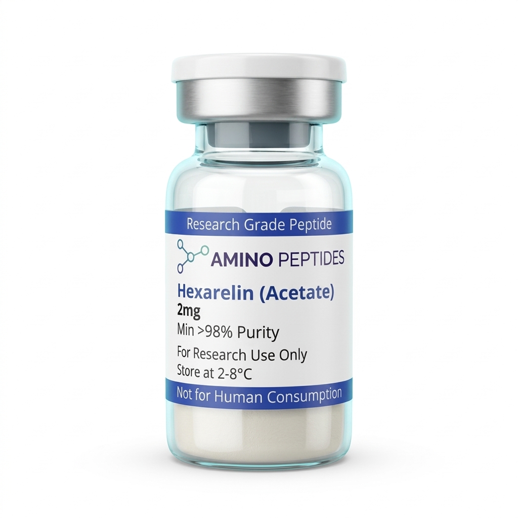
Hexarelin
From £13
Supplied as a sealed freeze-dried vial (stable 12–24 months at 2°C–8°C in a standard refrigerator).
Shop Hexarelin →What Preclinical Timelines Show from Early Signalling to Tissue Remodelling
When researchers set up an experiment with Hexarelin, the observed outcomes split cleanly into two phases: rapid biochemical signalling that happens within minutes, and structural tissue remodelling that takes weeks of consistent study protocols to measure.
In the first fifteen to sixty minutes after introducing a measured level of Hexarelin into a preclinical model, the primary readout is a sharp, clean spike in circulating growth hormone. Pituitary cells respond almost immediately to GHS-R1a activation by mobilizing intracellular calcium and releasing stored hormone granules. Alongside that primary wave, laboratory assays frequently record transient rises in prolactin and ACTH, which is a known trait of the strongest secretagogues compared to milder options like Ipamorelin. On the cardiovascular side, acute organ-bath studies show that within thirty minutes of low-oxygen stress, heart tissues exposed to Hexarelin maintain better left-ventricular developed pressure and release fewer damage markers such as lactate dehydrogenase and creatine kinase.
Moving past the first twenty-four to seventy-two hours, the focus shifts from immediate ion channel changes to gene expression and early repair signals. During this middle window, laboratory models display increased production of insulin-like growth factor 1 (IGF-1) in the liver as well as local IGF-1 splice variants inside muscle and vascular tissue. Endothelial nitric oxide synthase (eNOS) activity also begins to climb, which helps blood vessels relax and improves micro-circulation across stressed tissue beds.
The longer phase—typically spanning two to four weeks in rodent cardiovascular models—is where measurable structural changes appear. In published studies examining post-ischemic tissue remodelling, fourteen to twenty-eight days of protocol exposure resulted in less fibrotic scar formation in the ventricular wall. Instead of stiff, non-beating collagen fibres taking over the damaged zone, researchers observed better preservation of healthy cardiomyocyte alignment and improved capillary density around the border zone. One key timing detail noted in the literature is receptor desensitisation: when high concentrations of Hexarelin are applied nonstop for several weeks, the pituitary GH pulse can taper down by thirty to fifty percent, whereas the direct CD36-mediated cardiac effects appear far less prone to down-regulation. For that reason, many laboratory timelines use intermittent schedules or cycling windows to keep both pathways responsive across a full month of observation.
Comparison of Growth Hormone Secretagogues in Laboratory Models
| Peptide | Amino Acid Length | Primary Receptor Targets | GH Pulse Potency | Cortisol & Prolactin Spillover | Direct Cardiac CD36 Binding |
|---|---|---|---|---|---|
| Hexarelin | 6 amino acids (methylated) | GHS-R1a and CD36 | Very High | Moderate to High | Strong, well-characterised |
| GHRP-6 | 6 amino acids | GHS-R1a (weak CD36) | High | Moderate | Moderate |
| GHRP-2 | 6 amino acids | GHS-R1a | High | Low to Moderate | Minimal |
| Ipamorelin | 5 amino acids | GHS-R1a (highly selective) | Moderate | Negligible | None observed |
| Endogenous Ghrelin | 28 amino acids | GHS-R1a | Baseline reference | Low | Minimal (prefers GHS-R1a) |
Pairing Hexarelin with GHRHs and Vascular Peptides in Laboratory Stacks
In laboratory design, Hexarelin is rarely studied in complete isolation once its baseline metrics are known. Because it operates through the ghrelin receptor and the CD36 scavenger pathway, researchers frequently pair it with compounds that hit complementary receptors to see how multiple repair signals interact without competing for the same binding site.
The most classic pairing in endocrine and tissue recovery research combines Hexarelin with a growth hormone-releasing hormone (GHRH) analogue such as Mod GRF 1-29 (often called CJC-1295 without DAC) or Sermorelin. Here is why that combination is so widely tested: GHRH analogues bind to the GHRH receptor on pituitary somatotrophs and raise cyclic AMP (cAMP) levels, which tells the cell to produce more growth hormone. Hexarelin binds to the separate GHS-R1a receptor, raising phospholipase C and intracellular calcium to push that hormone out of the cell, while also lowering somatostatin—the brake pedal that normally stops release. When both peptides are present in a protocol ratio, the resulting hormone pulse recorded in published studies is significantly larger than adding the two separate effects together.
When the research goal leans heavily toward cardiovascular repair and blood vessel formation, scientists often look at pairing Hexarelin with BPC-157 or TB-500 (a synthetic fragment of Thymosin Beta-4). While Hexarelin guards cardiomyocytes against oxidative cell death and modulates fatty acid transport via CD36, BPC-157 upregulates vascular endothelial growth factor (VEGF) receptors to encourage new blood vessel sprouts. Meanwhile, TB-500 binds to actin monomers to help endothelial and smooth muscle cells migrate into damaged areas. Studying them side by side allows investigators to watch how cellular survival, actin migration, and capillary sprouting coordinate during tissue remodelling.
Another growing area of interest involves combining Hexarelin with mitochondrial support peptides such as SS-31 (Elamipretide) or MOTS-c in cellular energy assays. Because Hexarelin helps prevent mitochondrial membrane collapse from the outside via CD36 signalling, pairing it with a peptide that stabilises cardiolipin on the inner mitochondrial membrane gives researchers a two-layer model for examining ATP output under heavy metabolic stress.

Figure: Hexarelin: The Most Potent GHS for Cardiovascular Research?
Reconstitution calculator
Choose a vial mass and a bacteriostatic reconstitution solution volume. The figure is the resulting concentration.
Concentration: 2.50 mg/mL
Per 0.01mL (10µL): 25.0 mcg
Open the full calculatorBacteriostatic reconstitution solutionWhy 99% HPLC Purity and Cold Storage Matter for Hexarelin Vials
Even the best-designed cardiovascular assay will produce muddy, unreliable numbers if the peptide inside the vial is degraded or contaminated with synthesis leftovers. Because Hexarelin is a compact hexapeptide containing a modified D-2-methyl-tryptophan residue, verifying its chemical cleanliness before running an experiment is basic good housekeeping for any laboratory.
The first benchmark to check on a Certificate of Analysis (CoA) is High-Performance Liquid Chromatography (HPLC) purity at 99% or higher, paired with Mass Spectrometry (MS) confirmation of the exact molecular weight (887.04 Daltons). When a batch falls below 98% purity, the remaining percentage is not harmless filler; it usually consists of truncated peptide chains—five-amino-acid fragments missing a residue—or sequences where the protective chemical groups failed to detach cleanly. Those broken fragments can still partially occupy GHS-R1a or CD36 receptors without flipping the switch, acting like a key snapped off in a lock and dulling the measured cellular response.
Equally important for sensitive heart and endothelial cell cultures is trifluoroacetic acid (TFA) removal. Standard solid-phase synthesis uses TFA to cleave the finished peptide from its resin, leaving behind TFA salt counter-ions. In isolated cardiomyocyte or endothelial dishes, excess residual TFA shifts the local pH and causes direct cytotoxicity, killing cells or triggering stress pathways that have nothing to do with Hexarelin itself. High-grade research batches undergo ion-exchange chromatography to swap harsh TFA ions for a gentle acetate salt, keeping cellular assays clean and reproducible.
Once a verified lyophilised (freeze-dried) vial arrives, temperature discipline keeps the sequence intact. In its dry powder form, storing the vial in a freezer at -20°C locks the amino acid chain in place for long-term archiving. After adding a sterile bacteriostatic reconstitution solution—gliding the liquid slowly down the inside glass wall rather than squirting it straight onto the powder cake, and swirling gently instead of shaking—the reconstituted vial belongs in a dedicated refrigerator kept strictly between 2°C and 8°C. Leaving a liquid peptide at room temperature or exposing it to repeated freeze-thaw cycles breaks the peptide bonds and oxidises the tryptophan ring, turning an active reference standard into inactive fragments within days.
Frequently Asked Questions
Why is Hexarelin: The Most Potent GHS for Cardiovascular Research?
Researchers often ask if Hexarelin is the most potent growth hormone secretagogue for cardiovascular research because it binds strongly to cardiac CD36 and GHS-R1a receptors. Published studies show this dual signalling supports tissue remodelling and protects heart cells in laboratory models, making it a primary focus for ongoing cellular investigations.
How long do Hexarelin study timelines last in preclinical models?
Most laboratory investigations evaluating Hexarelin run for two to four weeks before receptor desensitisation occurs. In published studies using rodent models, researchers carefully track cellular signalling across these short intervals to observe tissue recovery and cardiac dynamics while avoiding the diminishing responses seen during extended exposure periods.
Can researchers combine Hexarelin with GHRH peptides in laboratory studies?
Yes, scientists frequently pair Hexarelin with growth hormone-releasing hormone analogues like Mod GRF 1-29 in laboratory models. Published studies demonstrate that combining these distinct compounds creates a synergistic signalling pulse, allowing researchers to analyse amplified endocrine pathways and characterise receptor interactions more effectively than testing a single compound alone.
How should Hexarelin be stored to maintain high purity for laboratory analysis?
Lyophilised Hexarelin must be kept in a freezer at minus 20 degrees Celsius to preserve its structural integrity and 99 percent purity. Once mixed with a bacteriostatic reconstitution solution for in vitro experiments, the liquid vial requires dark fridge storage between 2 and 8 degrees Celsius for up to four weeks.
Bibliography
- Mao Y, Tokudome T, Kishimoto I. The cardiovascular actions of hexarelin. Journal of Geriatric Cardiology. 2014;11(3):253-258. View published research
- Locatelli V, Rossoni G, Schweiger F, et al. Growth hormone-independent cardioprotective effects of hexarelin in the rat. Endocrinology. 1999;140(9):4024-4031. View published research
- Bodart V, Febbraio M, Demers A, et al. CD36 mediates the cardiovascular action of growth hormone-releasing peptides in the heart. Circulation Research. 2002;90(8):844-849. View published research
- Bisi G, Podio V, Valetto MR, et al. Acute cardiovascular and hormonal effects of GH and hexarelin, a synthetic GH-releasing peptide, in humans. Journal of Endocrinological Investigation. 1999;22(4):266-272. View published research
- degli Uberti EC, Ambrosio MR, Bondanelli M, et al. Human administration of hexarelin, a synthetic growth hormone-releasing peptide, induces both growth hormone and adrenocorticotropin/cortisol release. Journal of preclinical Endocrinology & Metabolism. 1995;80(10):2943-2948. View published research
See our UK peptide research guides & COA briefings first in Google Search.
Add Amino Peptides UK as a Preferred Source on Google⚠️ Research Use Only Disclaimer: All peptides and compounds are sold strictly for in-vitro laboratory research purposes only. Not intended for human or veterinary use, not a dietary supplement, and not approved to diagnose, treat, cure, or prevent any disease or condition. For use by qualified researchers in suitably equipped laboratory environments only. Amino Peptides Ltd operates in full compliance with UK MHRA regulations and applicable EU research chemical guidelines.