
IGF-1 LR3: The Impact of Long R3 Analogues on Muscle Cell Hyperplasia
14th Aug 2026
What You Need to Know About IGF-1 LR3 Peptide
- Researchers investigate IGF-1 LR3 to understand how modified growth factors drive the creation of brand-new muscle cells, a process known as hyperplasia, rather than merely expanding existing cell size.
- The molecule works by bypassing natural binding proteins in the bloodstream, allowing it to stay active much longer and trigger cellular division pathways inside skeletal muscle tissue.
- Published studies in preclinical models show that this structural change leads to roughly three times the biological potency of native insulin-like growth factor alongside a significantly extended active window.
- Because it binds poorly to carrier proteins, laboratory models demonstrate rapid nutrient shuttling and intense satellite cell proliferation without immediate clearance by the body.
How the Long R3 Modification Unlocks Satellite Cell Division
IGF-1 LR3 is an eighty-three amino acid synthetic analogue of Insulin-like Growth Factor-1 that substitutes arginine for glutamic acid at position three and adds a thirteen-amino-acid extension at the N-terminus. In published studies, this specific modification blocks carrier proteins from trapping the peptide, leaving it free to trigger satellite cell division and muscle cell hyperplasia in laboratory models.
To understand why researchers study IGF-1 LR3: The Impact of Long R3 Analogues on Muscle Cell Hyperplasia, it helps to first separate two basic concepts: hypertrophy and hyperplasia. Hypertrophy is when existing muscle cells simply swell and grow larger, much like inflating a small number of balloons to their maximum size. Hyperplasia is the actual splitting and multiplication of cells to create entirely new muscle fibres, increasing the total number of cells in the tissue.
In native biology, standard IGF-1 consists of seventy amino acids. The moment it enters a biological system, sticky carrier proteins called Insulin-like Growth Factor Binding Proteins (IGFBPs) grab onto it. Up to ninety-eight percent of native IGF-1 gets locked up by these binding proteins, which leaves it inactive and gives the remaining free fraction a very short active life of less than twenty minutes.
Biochemists designed the Long R3 analogue to bypass that trap. By swapping the third amino acid from glutamic acid to arginine (the R3 change) and attaching a thirteen-amino-acid tail to the front of the chain (the Long extension), the molecule changes shape just enough that IGFBPs can no longer grip it tightly. Its affinity for those inhibitory binding proteins drops by more than a hundredfold, while its ability to dock with the Type 1 IGF receptor on muscle membranes stays intact.
Because it stays free in the surrounding fluid for twenty to thirty hours in preclinical models, IGF-1 LR3 keeps knocking on the cell door. This extended contact wakes up dormant stem-like cells sitting on the outside of muscle fibres, known as satellite cells. Once activated through the PI3K/Akt and MAPK intracellular signalling pathways, these satellite cells wake up, multiply, and fuse into the tissue to form fresh myoblasts and new structural fibres, driving true cellular hyperplasia alongside tissue recovery and nitrogen retention.
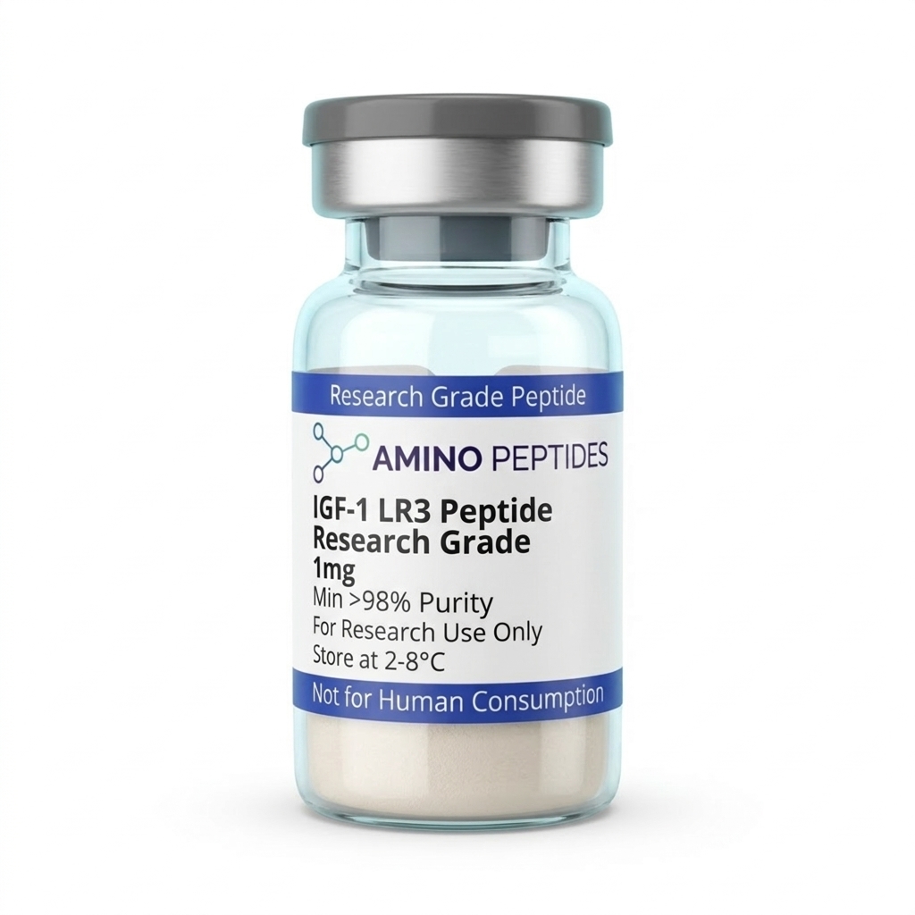
IGF-1 LR3 Peptide
From £59
Supplied as a sealed freeze-dried vial (stable 12–24 months at 2°C–8°C in a standard refrigerator).
Shop IGF-1 LR3 Peptide →What Preclinical Timelines Show From Early Signalling to Fibre Splitting
When scientists observe IGF-1 LR3 in cell cultures and preclinical rodent models, the biological response unfolds in distinct stages. Because hyperplasia requires a dormant stem cell to wake up, copy its DNA, divide, and mature into a working muscle fibre, the timeline looks very different from a simple fluid shift or temporary metabolic spike.
During the first one to twenty-four hours of exposure in laboratory dishes, the activity is purely chemical. Within minutes of introducing a measured level of IGF-1 LR3 to cultured myoblasts, the peptide binds to transmembrane Type 1 IGF receptors. This triggers immediate phosphorylation of insulin receptor substrate-1 (IRS-1) and switches on the Akt/mTOR pathway. During this initial twenty-four-hour window, researchers measure a sharp rise in glucose transport into the cells and a rapid increase in amino acid uptake, preparing the cellular environment with the raw materials needed for division.
Between days two and seven in published studies, satellite cell proliferation begins in earnest. The MAPK/ERK signalling pathway pushes quiescent satellite cells out of their resting state (the G0 phase) and into active mitosis. Under a microscope, researchers track a clear climb in the number of myonuclei per millimetre of fibre. Instead of just making more protein inside old walls, the tissue is actively building new nuclear control centres to support future fibres.
By weeks two through four in longer preclinical models, those newly divided myoblasts line up, fuse together, and form young myotubes. This is the stage where structural tissue remodelling becomes visible on histological cross-sections. Laboratory analyses show a larger total count of smaller-diameter, newly formed muscle fibres sitting beside mature fibres—the classic signature of hyperplasia.
One critical detail observed across multi-week study timelines is receptor downregulation. Because IGF-1 LR3 stays active for roughly twenty to thirty hours and resists clearance by binding proteins, continuous high concentrations eventually cause the target cells to pull their Type 1 IGF receptors inside the membrane to rest. For this reason, published laboratory protocols typically limit continuous exposure windows to four to six weeks, followed by an equal washout period so membrane receptor density can return to baseline.
Biochemical Comparison: Native IGF-1 vs. IGF-1 LR3 vs. DES (1-3) IGF-1
| Characteristic | Native IGF-1 | IGF-1 LR3 (Long R3) | DES (1-3) IGF-1 |
|---|---|---|---|
| Amino Acid Length | 70 amino acids | 83 amino acids (13-residue extension) | 67 amino acids (missing first 3 residues) |
| Key Structural Feature | Glutamic acid at position 3 | Arginine substitution at position 3 plus N-terminal leader sequence | Truncated N-terminus removing the primary binding site |
| IGFBP Binding Affinity | High (nearly 99 percent bound in circulation) | Very low (over 100-fold reduction in binding affinity) | Very low (bypasses local tissue binding proteins) |
| Active Half-Life in Models | Approximately 10 to 20 minutes (unbound) | Approximately 20 to 30 hours | Approximately 20 to 30 minutes |
| Primary Research Focus | Baseline endocrine signalling and systemic growth regulation | Sustained systemic satellite cell proliferation and hyperplasia | Rapid, localised tissue remodelling and site-specific receptor activation |
Pairing IGF-1 LR3 With Complementary Research Peptides in the Lab
In laboratory research, investigators rarely look at a single signalling pathway in isolation. Muscle regeneration and hyperplasia involve multiple steps: waking up dormant satellite cells, instructing them to multiply, and then feeding those new cells enough structural protein to mature into full fibres. Because each peptide family targets a different step in this chain, researchers frequently study IGF-1 LR3 alongside complementary compounds.
The most common pairing in published literature combines IGF-1 LR3 with Mechano Growth Factor (MGF) or its stabilised version, PEG-MGF. While both molecules come from the same parent gene family, they act at different times during tissue remodelling. In preclinical models of mechanical strain, MGF acts as the initial spark that replenishes the pool of raw satellite stem cells, stopping them from maturing too early so more of them can multiply. Once MGF has expanded that stem cell pool, IGF-1 LR3 steps in to drive those cells through final differentiation and fusion into new muscle fibres. Studying them in sequence allows researchers to observe both pool expansion and fibre maturation without the two signals competing for the same receptor at the exact same hour.
Another widely studied pairing involves growth hormone secretagogues such as CJC-1295 and Ipamorelin. In isolated models, a high concentration of exogenous IGF-1 LR3 can signal the pituitary gland to slow down its own natural pulsatile output through negative feedback. By pairing a secretagogue combination like CJC-1295 and Ipamorelin alongside an IGF-1 LR3 protocol ratio, researchers investigate how maintaining natural somatotropic pulses supports fat oxidation and extracellular matrix repair while the Long R3 analogue drives local satellite cell hyperplasia.
For protocols focused on complete structural recovery, investigators also pair IGF-1 LR3 with BPC-157 and TB-500. When muscle tissue splits and forms new fibres through hyperplasia, it needs a matching network of fresh blood vessels and connective tissue fascia to hold the new cells and supply them with oxygen. BPC-157 and TB-500 promote angiogenesis and fibroblast migration, ensuring the surrounding scaffolding keeps pace with the multiplying muscle cells. Meanwhile, teams investigating neuromuscular junction signalling alongside muscle remodelling often cross-reference cognitive research peptides to study how central neural drive and peripheral tissue adaptation interact.

Figure: IGF-1 LR3: The Impact of Long R3 Analogues on Muscle Cell Hyperplasia
Reconstitution calculator
Choose a vial mass and a bacteriostatic reconstitution solution volume. The figure is the resulting concentration.
Concentration: 2.50 mg/mL
Per 0.01mL (10µL): 25.0 mcg
Open the full calculatorBacteriostatic reconstitution solutionWhy 99%+ Purity, Acetic Acid Reconstitution, and Cold Storage Matter
Working with an eighty-three amino acid polypeptide like IGF-1 LR3 requires much stricter quality standards than handling short five- or ten-amino-acid chains. Every extra amino acid link added during solid-phase synthesis introduces another opportunity for incomplete coupling, deletion sequences, or misfolded chains. If a batch stops at ninety-two or ninety-five percent purity, the remaining five to eight percent is not harmless filler—it is a soup of truncated peptide fragments and toxic synthesis leftovers.
Truncated fragments are especially disruptive in hyperplasia research. A broken seventy-amino-acid fragment might still bind to the Type 1 IGF receptor on the outside of a muscle cell without flipping the switch inside, acting as a dead-weight blocker that stops intact IGF-1 LR3 molecules from working. Even worse, residual Trifluoroacetic Acid (TFA) left behind from cheap cleavage processes lowers cellular pH and damages delicate myoblast cultures on contact. Verifying that every vial meets a 99%+ purity threshold via High-Performance Liquid Chromatography (HPLC) and Mass Spectrometry (MS), with thorough TFA removal, ensures that the biological response observed in the lab comes strictly from intact IGF-1 LR3.
Reconstitution chemistry is another major factor that separates reliable IGF-1 LR3 data from failed experiments. Unlike smaller peptides that dissolve effortlessly in standard solutions, the Long R3 chain carries an isoelectric point and hydrophobic profile that can cause it to clump, cloud up, or stick to the glass walls of the vial if mixed in neutral pH liquids alone. In laboratory protocols, researchers reconstitute lyophilised IGF-1 LR3 using sterile diluted acetic acid (typically zero point six percent) to gently lower the pH, fully dissolve the eighty-three amino acid chain without aggregation, and preserve its folded shape over multi-week study timelines.
Once reconstituted, temperature control determines how long that chain stays intact. Lyophilised powder remains stable in freezer storage at minus twenty degrees Celsius for long periods, but liquid IGF-1 LR3 must be kept strictly in a refrigerator between 2°C and 8°C. Gently swirling the vial down the glass wall—never shaking it violently—protects the delicate tertiary bridges that allow the peptide to trigger satellite cell hyperplasia.
Frequently Asked Questions
Why do researchers study IGF-1 LR3 for muscle cell hyperplasia?
Researchers examine this modified peptide because it resists binding proteins and stays active much longer than natural IGF-1 in laboratory models. Published studies show it stimulates satellite cells to divide and form brand-new muscle fibres, allowing scientists to analyse cellular growth, tissue remodelling, and structural regeneration in isolated environments.
How long do laboratory studies on IGF-1 LR3 typically last?
Most preclinical models observe the effects of this analogue over a four to six week period. Because continuous exposure can cause cellular receptors to become less responsive over time, researchers carefully schedule their protocol ratios and study amounts to characterise peak signalling before receptor sensitivity naturally drops off.
Do scientists combine IGF-1 LR3 with other peptides in research?
Yes, laboratory models frequently pair IGF-1 LR3 with growth hormone secretagogues or mechanical growth factors to observe overlapping signalling pathways. Studying these combinations helps scientists understand how different amino acid chains interact to optimise cellular recovery, protein synthesis, and satellite cell activation during controlled tissue regeneration experiments.
How is IGF-1 LR3 stored to maintain high purity in the lab?
Lyophilised IGF-1 LR3 powder must be kept in a dark laboratory freezer at minus twenty degrees Celsius to prevent degradation. Once mixed with a sterile acetic acid solution or bacteriostatic reconstitution solution, the liquid sample should be stored in a standard refrigerator and utilised within three to four weeks.
Bibliography
- Francis GL, Ross M, Ballard FJ, et al. Novel recombinant fusion protein analogues of insulin-like growth factor (IGF)-I indicate the relative importance of IGF-binding protein and receptor binding for enhanced biological potency. J Mol Endocrinol. 1992;8(3):213-223. View published research
- Tomas FM, Knowles SE, Owens PC, et al. Effects of full-length and truncated insulin-like growth factor-I on nitrogen balance and muscle protein metabolism in nitrogen-restricted rats. J Endocrinol. 1992;132(3):397-405. View published research
- Barton-Davis ER, Shoturma DI, Musaro A, Rosenthal N, Sweeney HL. Viral mediated expression of insulin-like growth factor I blocks the aging-related loss of skeletal muscle function. Proc Natl Acad Sci U S A. 1998;95(26):15603-15607. View published research
- Velloso CP. Regulation of muscle mass by growth hormone and IGF-I. Br J Pharmacol. 2008;154(3):557-568. View published research
- Adams GR. Role of insulin-like growth factor-I in the regulation of skeletal muscle adaptation to increased loading. Exerc Sport Sci Rev. 1998;26:31-60. View published research
⚠️ Research Use Only Disclaimer: All peptides and compounds are sold strictly for in-vitro laboratory research purposes only. Not intended for human or veterinary use, not a dietary supplement, and not approved to diagnose, treat, cure, or prevent any disease or condition. For use by qualified researchers in suitably equipped laboratory environments only. Amino Peptides Ltd operates in full compliance with UK MHRA regulations and applicable EU research chemical guidelines.