
The IGF-1 LR3 Trend Analysed: Fact vs Fiction, Cellular Mechanisms, and Long-Term Safety
6th Nov 2026
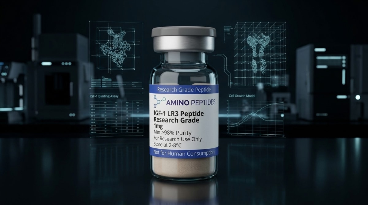
What You Need to Know About IGF-1 LR3 Peptide
- Online fitness communities frequently discuss this compound for rapid muscle tissue remodelling, though internet anecdotes often ignore severe metabolic risks.
- The molecule works by unlocking surface receptors on cells to trigger internal cascades that boost amino acid uptake and block protein breakdown.
- Because it is engineered to bypass natural carrier proteins, it stays active in laboratory models up to twenty times longer than native growth factor.
- Published studies in animal and cell models show strong tissue regeneration alongside concerning blood sugar drops and unwanted organ enlargement.
How Long-R3 IGF-1 Sidesteps Binding Proteins to Drive Cellular Recovery
Long-R3 Insulin-like Growth Factor-1 (IGF-1 LR3) is an engineered eighty-three-amino-acid polypeptide designed to resist deactivation by insulin-like growth factor binding proteins in cell culture. By swapping a glutamic acid for an arginine at position three and adding a thirteen-amino-acid extension at the N-terminus, the molecule stays active in solution far longer than native IGF-1.
In standard biological systems, native IGF-1 consists of seventy amino acids. Almost immediately after secretion, roughly ninety-eight percent of native IGF-1 gets trapped by a family of six carrier molecules called Insulin-like Growth Factor Binding Proteins (IGFBPs). While these binding proteins extend the circulation time of the native hormone, they physically block it from docking with the IGF-1 receptor (IGF-1R) on the surface of target cells. For researchers studying tissue remodelling and cellular energy in isolated myoblasts or fibroblasts, this natural braking system limits how much direct receptor signalling can be observed at a given study amount.
Biochemists developed the LR3 variant specifically to bypass that bottleneck in laboratory models. The substitution of arginine (R) for glutamic acid (E) at the third position of the amino acid chain, combined with the thirteen-residue N-terminal extension peptide, changes the three-dimensional charge distribution of the molecule. Because of this structural tweak, IGFBPs have a much harder time grabbing onto IGF-1 LR3. Published studies show its affinity for binding proteins is reduced by more than a hundredfold compared to native IGF-1, leaving the vast majority of the peptide free in the surrounding medium to interact directly with cell-surface receptors.
Once free IGF-1 LR3 docks with the transmembrane IGF-1R, it triggers autophosphorylation of the receptor's intracellular tyrosine kinase domains. This action switches on two primary internal pathways: the phosphoinositide 3-kinase (PI3K/Akt) pathway and the mitogen-activated protein kinase (MAPK/ERK) pathway. In preclinical models of skeletal muscle satellite cells, activating Akt promotes glucose transport across the cell membrane, accelerates amino acid uptake, and switches on mammalian target of rapamycin (mTOR) signalling to build new structural proteins. At the same time, the ERK pathway signals dormant stem-like satellite cells to divide and fuse with existing fibres, supporting structural recovery and hypertrophy in the dish.
Online forum discussions — including frequent threads on Reddit — often blur the line between these isolated cell-culture observations and whole-body outcomes. When curious readers search for daily protocol ratios or expected outcomes, they rarely see mention of the fact that IGF-1R is present on almost every tissue type, not just skeletal muscle. In preclinical models, unbuffered activation of these receptors across multiple organ systems explains why researchers monitor closely for off-target proliferation and rapid drops in extracellular glucose levels.
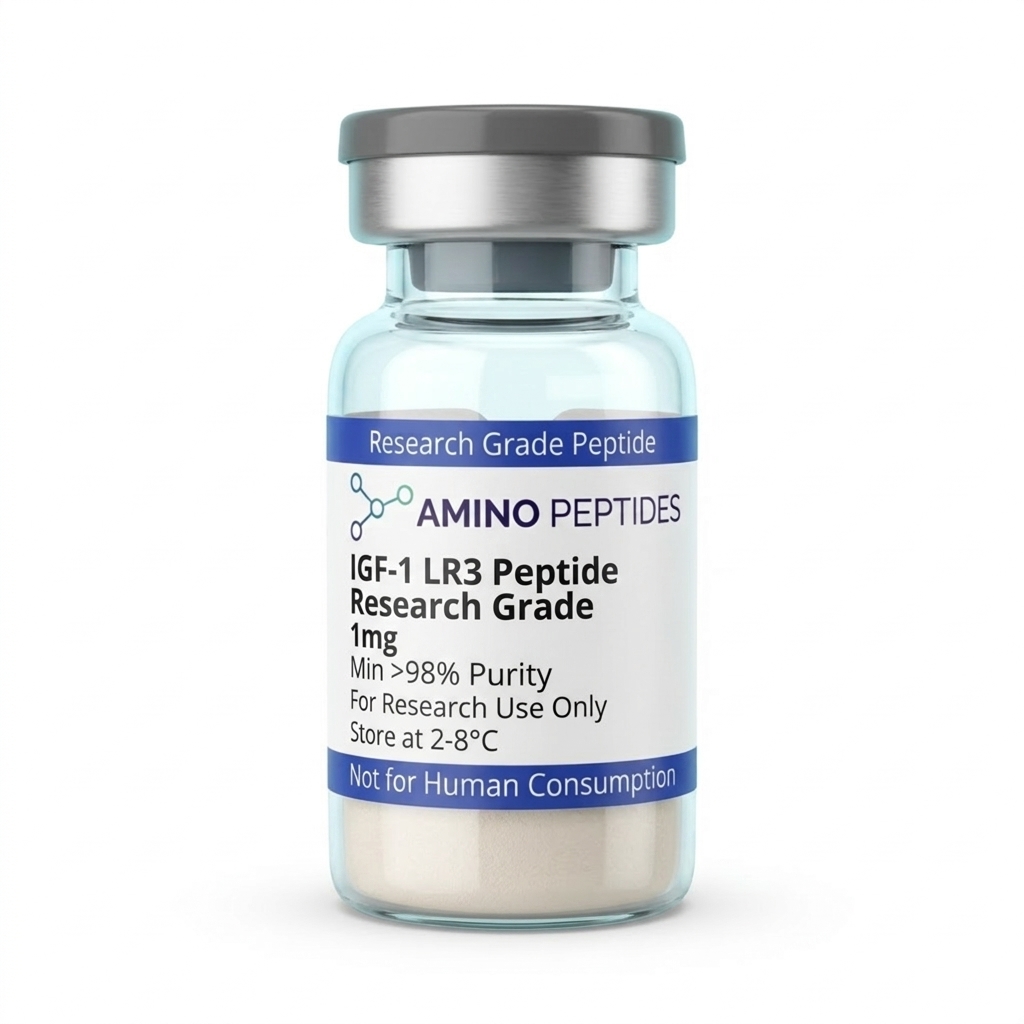
IGF-1 LR3 Peptide
From £59
Supplied as a sealed freeze-dried vial (stable 12–24 months at 2°C–8°C in a standard refrigerator).
View published researchWhat Preclinical Timelines Show About Early Signalling and Receptor Desensitisation
Tracking the activity of IGF-1 LR3 in laboratory models requires separating immediate biochemical reactions from slower structural remodelling. Because the modified peptide avoids capture by IGFBPs, its active half-life in biological fluids extends to roughly twenty to thirty hours, compared to less than fifteen minutes for unbound native IGF-1. This extended window changes how quickly and how long target cells respond after a single measured level is introduced to the culture medium.
During the first fifteen to sixty minutes of exposure in vitro, researchers observe rapid phosphorylation of the IGF-1 receptor and insulin receptor substrate-1 (IRS-1). Within two to four hours, GLUT4 transporters migrate to the cell membrane, pulling glucose out of the surrounding medium to fuel cellular energy production. By the twelve-to-twenty-four-hour mark, ribosomal protein synthesis rates climb measurably, and gene expression markers associated with cell cycle progression — such as cyclin D1 — begin to peak in cultured myoblasts and fibroblasts.
Structural tissue remodelling in preclinical models takes considerably longer to appear. Over a three-to-seven-day window of controlled exposure, satellite cells complete multiple rounds of mitotic division and begin differentiating into mature myotubes. By days ten to fourteen, histological cross-sections in animal studies show increased myofibrillar density, enhanced collagen matrix deposition in connective tissues, and measurable changes in local nitrogen retention. These structural shifts depend heavily on the concentration used and the nutrient density of the surrounding environment.
However, longer timelines in published studies also highlight a built-in biological limit: receptor downregulation and desensitisation. When cells are exposed to high measured levels of IGF-1 LR3 continuously over several weeks, they protect themselves from overstimulation by pulling IGF-1R proteins inside the cell membrane and degrading them via lysosomes. In preclinical models, continuous exposure beyond three to four weeks often produces diminishing returns in protein synthesis as receptor density drops. Furthermore, long-term safety evaluations in animal models note that sustained, high-concentration exposure can stimulate unwanted growth in visceral organs, alter local insulin sensitivity due to cross-talk with insulin receptors, and stress metabolic feedback loops. For this reason, published laboratory protocols typically limit observation periods to short, intermittent windows rather than continuous long-term runs.
Biochemical Comparison: Native IGF-1 vs Modified Laboratory Analogues
| Feature | Native IGF-1 | IGF-1 LR3 | IGF-1 DES (1-3) |
|---|---|---|---|
| Amino Acid Length | 70 amino acids | 83 amino acids (includes 13-residue extension) | 67 amino acids (missing first 3 residues) |
| Key Structural Modification | Unmodified endogenous sequence | Glutamic acid replaced by Arginine at position 3, plus N-terminal tail | Cleavage of N-terminal tripeptide (Gly-Pro-Glu) |
| IGFBP Binding Affinity | High (over 98% bound to carrier proteins in circulation) | Very low (engineered to evade binding proteins) | Low (reduced affinity due to truncated N-terminus) |
| Approximate Clearance Rate | Rapid for free form (10 to 20 minutes); hours when bound | Extended (20 to 30 hours in laboratory models) | Very rapid (minutes, acts primarily at local tissue sites) |
| Primary Research Context | Baseline endocrine signalling and metabolic homeostasis | Bioreactor cell culture productivity and systemic growth models | Localised tissue repair and neurobiology models |
Pairing Growth Factor Analogues with Complementary Peptides in Lab Models
In academic and commercial laboratories, investigators rarely look at growth factors in complete isolation. Because tissue regeneration and cellular maintenance involve multiple overlapping signals, researchers frequently design experiments that pair IGF-1 LR3 with complementary peptides to see how different pathways interact without overloading a single receptor type.
One common experimental pairing combines IGF-1 LR3 with Mechano Growth Factor (MGF) or its pegylated analogue, PEG-MGF. While both molecules originate from the same parent gene in mammals, they perform distinct jobs during tissue remodelling. In preclinical muscle recovery models, MGF acts as the initial spark that wakes up quiescent satellite cells and expands the local stem cell pool, while delaying their final differentiation. Once that pool of precursor cells is expanded, IGF-1 LR3 drives them to fuse with existing fibres and synthesise new contractile proteins. Studying the two in sequence allows researchers to observe how precursor proliferation and mature hypertrophy balance each other.
Another frequent research focus involves combining IGF-1 LR3 with cytoprotective and angiogenic peptides such as BPC-157 and TB-500 (a synthetic fragment of Thymosin Beta-4). While IGF-1 LR3 supplies the anabolic signal for protein assembly via the PI3K/Akt pathway, rapid tissue growth in three-dimensional cultures or animal models requires an adequate blood supply and an organised extracellular matrix. BPC-157 upregulates vascular endothelial growth factor (VEGF) expression to support new capillary formation, while TB-500 binds to globular actin to help endothelial cells and fibroblasts migrate to the site of remodelling. Together, these pathways allow investigators to study how vascular support and structural protein synthesis coordinate during recovery.
Researchers exploring broader cellular resilience and central nervous system signalling also compare peripheral growth factors alongside neuropeptide classes such as Semax and Selank, which modulate brain-derived neurotrophic factor (BDNF) and GABAergic signalling in preclinical models. Comparing how peripheral tyrosine kinase receptors and central neurotrophic pathways respond to stress gives biochemists a clearer view of whole-organism adaptation. When teams buy igf-1 lr3 for multi-peptide assays, keeping each compound's concentration within physiological study bounds prevents receptor saturation and keeps downstream data clean.
Figure 1: The IGF-1 LR3 Trend Analysed: Fact vs Fiction, Cellular Mechanisms, and Long-Term Safety
Reconstitution calculator
Choose a vial mass and a bacteriostatic reconstitution solution volume. The figure is the resulting concentration.
Concentration: 2.50 mg/mL
Per 0.01mL (10µL): 25.0 mcg
Open the full calculatorBacteriostatic reconstitution solutionWhy 99% HPLC Purity and Cold Storage Matter for an 83-Amino-Acid Chain
Working with an eighty-three-amino-acid polypeptide presents very different analytical challenges than working with short tripeptides or pentapeptides. As a synthetic chain grows longer on a solid-phase resin, the chance of incomplete amino acid coupling, deletion sequences, or misfolded disulphide bridges rises sharply. IGF-1 LR3 contains three internal disulphide bonds that must fold into a specific tertiary shape so the molecule can fit into the IGF-1 receptor binding pocket. If a batch contains truncated sequences or misfolded isomers, those flawed molecules can compete for receptor space without triggering full intracellular signalling, skewing every assay readout.
To avoid this problem, laboratory-grade IGF-1 LR3 must be verified at ninety-nine percent or higher purity using High-Performance Liquid Chromatography (HPLC) alongside Mass Spectrometry (MS) to confirm the exact molecular weight of 9,111 Daltons. During solid-phase synthesis, trifluoroacetic acid (TFA) is routinely used to cleave the finished peptide from its resin and remove protective side-chain groups. If residual TFA salts are not thoroughly exchanged out during freeze-drying, they lower the pH of the reconstituted solution and prove toxic to delicate cell cultures, causing membrane damage that looks like a side effect of the peptide itself. Clean, counter-ion-exchanged lyophilised powder protects cell viability and keeps experimental baselines steady.
Because of its folded three-dimensional structure, IGF-1 LR3 is sensitive to heat, mechanical agitation, and repeated freeze-thaw cycles. Prior to reconstitution, the lyophilised powder should be stored at minus twenty degrees Celsius away from light. When preparing the vial for laboratory use, researchers introduce the solvent — such as dilute acetic acid or a suitable bacteriostatic reconstitution solution depending on the specific assay protocol — slowly down the inside glass wall rather than squirting it directly onto the powder cake. Vigorous shaking can shear the disulphide bridges and cause the protein to clump together into inactive aggregates; gentle swirling is always used instead. Once in liquid form, the vial must be kept in a refrigerator between two and eight degrees Celsius and used within its validated stability window so oxidation and hydrolysis do not degrade the active sequence.
Frequently Asked Questions
Why are so many people searching for IGF-1 LR3 on Reddit and search engines?
Online curiosity about IGF-1 LR3 stems from its reputation in laboratory models for promoting rapid cellular growth and extended biological activity. Forum users frequently debate how its modified structure avoids binding proteins, which allows it to trigger prolonged tissue remodelling and muscle cell division in preclinical models.
What does a typical study timeline and measured level look like in published studies?
Published studies typically observe IGF-1 LR3 over short cycles of two to four weeks to monitor receptor sensitivity. Because this synthetic analogue stays active for roughly twenty hours, laboratory protocols evaluate daily study amounts in microgram concentrations to track cellular signalling without causing rapid receptor burnout.
Do laboratory protocols combine IGF-1 LR3 with other growth peptides?
Yes, preclinical models frequently pair IGF-1 LR3 with growth hormone secretagogues or PEG-MGF to analyse synergistic tissue recovery. Researchers observe how combining these distinct compounds influences satellite cell activation and protein synthesis pathways at different stages of cellular regeneration without overlapping their primary signalling mechanisms.
How is IGF-1 LR3 purity verified and how should it be stored in a laboratory?
Laboratory-grade IGF-1 LR3 requires third-party High-Performance Liquid Chromatography testing to confirm a purity level above ninety-eight percent. Before mixing with acetic acid or bacteriostatic reconstitution solution, freeze-dried vials are kept frozen, while mixed liquid solutions must stay refrigerated between two and eight degrees Celsius.
Bibliography
- Francis GL, Ross M, Ballard FJ, et al. Novel recombinant fusion protein analogues of insulin-like growth factor (IGF)-I indicate the relative importance of IGF-binding protein and receptor binding for enhanced biological potency. J Mol Endocrinol. 1992;8(3):213-223. View published research
- Tomas FM, Knowles SE, Owens PC, et al. Insulin-like growth factor-I (IGF-I) and especially IGF-I variants are anabolic in dexamethasone-exposed rats. Biochem J. 1992;282(Pt 1):91-97. View published research
- Laron Z. Insulin-like growth factor 1 (IGF-1): a growth hormone. Mol Pathol. 2001;54(5):311-316. View published research
- Philippou A, Maridaki M, Halapas A, Koutsilieris M. The role of the insulin-like growth factor 1 (IGF-1) in skeletal muscle physiology. In Vivo. 2007;21(1):45-54. View published research
- Pollak M. The insulin and insulin-like growth factor receptor family in neoplasia: an update. Nat Rev Cancer. 2012;12(3):159-169. View published research
⚠️ Research Use Only Disclaimer: All peptides and compounds are sold strictly for in-vitro laboratory research purposes only. Not intended for human or veterinary use, not a dietary supplement, and not approved to diagnose, treat, cure, or prevent any disease or condition. For use by qualified researchers in suitably equipped laboratory environments only. Amino Peptides Ltd operates in full compliance with UK MHRA regulations and applicable EU research chemical guidelines.