
IGF-1 LR3 vs. Native IGF-1: A Comparative Analysis of Bioavailability
21st Jun 2026

Quantifying cellular proliferation often requires precise isolation of the insulin-like growth factor pathway. While native Insulin-like Growth Factor 1 (IGF-1)—a highly conserved 70-amino-acid polypeptide—initiates these biochemical reactions, its viability in standard assay media is restricted by rapid degradation and a strong affinity for carrier proteins. Synthetic structural analogues, such as Long Arginine 3 IGF-1 (IGF-1 LR3), provide a functional alternative. This analysis evaluates the structural variances, binding dynamics, and half-life differences between these molecules under controlled in-vitro conditions.
Primary Structural and Kinetic Distinctions
- Structural Profile: IGF-1 LR3 features a 13-amino-acid N-terminal extension alongside a glutamic acid-to-arginine substitution at position 3.
- IGFBP Evasion: These engineered alterations significantly diminish binding affinity for insulin-like growth factor-binding proteins (IGFBPs).
- Media Half-Life: Evading IGFBP sequestration allows IGF-1 LR3 to maintain a prolonged active state within cellular culture systems compared to the native sequence.
- Free Bioavailability: Eliminating binding protein interference ensures greater concentrations of unbound peptide remain available to stimulate the IGF-1 receptor (IGF-1R).
Structural Alterations and Binding Dynamics
Native IGF-1 naturally binds to six high-affinity proteins, identified as IGFBPs. Within an isolated in-vitro environment, these proteins regulate activity by sequestering the peptide, which effectively blocks IGF-1R interaction. Although normal in complex biological systems, this sequestration prevents researchers from maintaining stable peptide gradients across culture media.
Synthesising the IGF-1 LR3 analogue necessitates replacing the position 3 glutamic acid residue with arginine (the 'R3' modification) and affixing a 13-amino-acid sequence to the N-terminus (the 'Long' modification). Modifying the tertiary structure and electrostatic profile decreases IGFBP binding affinity by over 120-fold. As a result, the peptide resists inhibitory attachment, ensuring a significantly larger free fraction remains available for assay interactions.
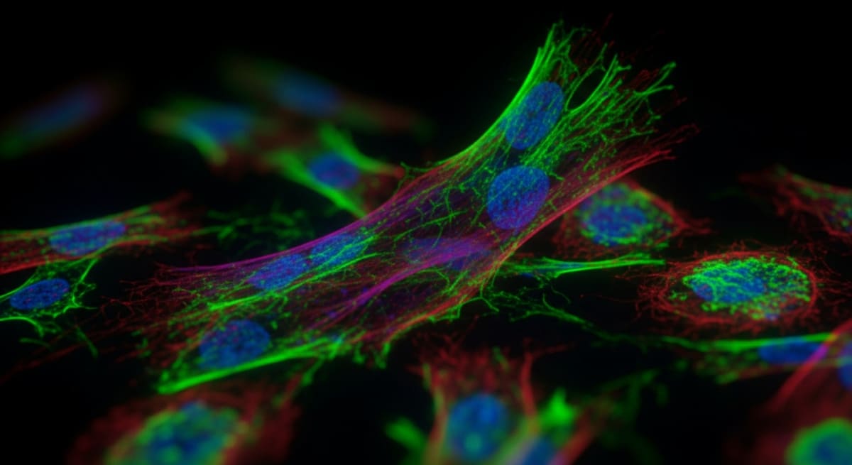
Figure 1: A high-resolution fluorescent microscopy view showing vibrant, glowing neon green, magenta, and cyan cellular structures against a pitch-black background.
Pharmacokinetics and Half-Life in Laboratory Models
Extended in-vitro assays depend heavily on peptide stability within the culture medium. Native IGF-1 exhibits a highly transient nature, yielding a half-life measured in minutes due to rapid enzymatic degradation. Binding to IGFBPs extends this half-life; however, the sequestered peptide remains biologically inactive.
Conversely, IGF-1 LR3 maintains an active half-life of 20 to 30 hours in laboratory media. Unbound and resistant to common degradation pathways, this extended stability permits investigators to sustain continuous receptor activation without frequent media changes. Sustained peptide presence is critical for measuring long-term cellular differentiation. Preserving structural integrity upon reconstitution requires sterile bacteriostatic diluents sourced from a reputable UK research supplier.
Receptor Activation Dynamics
Despite modifications designed to inhibit IGFBP attachment, IGF-1 LR3 retains a high affinity for its primary signalling target, IGF-1R. The essential binding domain required to dock with the extracellular alpha-subunits remains unaffected by the Glu3Arg substitution and N-terminal extension. Upon attachment, the analogue initiates autophosphorylation of the receptor's intracellular tyrosine kinase domain. This event subsequently triggers downstream phosphatidylinositol 3-kinase (PI3K)/Akt and mitogen-activated protein kinase (MAPK) signalling cascades in isolated cell cultures.
Because binding proteins function as buffers in unpurified media, achieving comparable receptor occupancy with native IGF-1 requires excessive peptide concentrations. Comparative in-vitro evaluations indicate that IGF-1 LR3 operates with greater efficiency, requiring lower molar concentrations to induce matching levels of receptor phosphorylation. This high efficiency establishes it as an accurate reagent for mapping growth kinetics, frequently evaluated alongside compounds like CJC-1295 or the cell-modulating TB-500 peptide.
Quality Verification and Analytical Testing
Laboratory controls necessitate rigorous verification of synthesised peptides prior to in-vitro application. Analysts deploy advanced spectroscopic and chromatographic methods to confirm purity, identity, and sequence integrity.
Function of the Peptide Certificate of Analysis
A peptide certificate of analysis provides empirical evidence documenting a synthetic batch's chemical identity and overall composition. The report supplies reversed-phase high-performance liquid chromatography (RP-HPLC) chromatograms to quantify purity, which frequently exceeds 98%. Mass spectrometry (MS) profiles, such as MALDI-TOF or ESI-MS, verify that the empirical molecular weight exactly corresponds to the target sequence's theoretical mass.
Verification by an Accredited Amino Acid Analysis Laboratory
Absolute quantification of the primary sequence requires testing by an accredited amino acid analysis laboratory. Protocols demand complete acid hydrolysis of the peptide backbone into free amino acids, followed by ion-exchange chromatography or reversed-phase HPLC separation. Post-column derivatisation, often using ninhydrin, allows analysts to confirm that the stoichiometric ratios of the recovered amino acids match the theoretical sequence.
Amino Acid Analysis Standards for UK Researchers
For investigators operating within Great Britain, sourcing verified amino acid analysis UK standards is essential for experimental standardisation. Leading suppliers provide a detailed certificate of analysis for peptides outlining both purity and net peptide content. This metric ensures molar concentrations are calculated accurately for cellular assays, free from distortions caused by counter-ions or moisture.
Interpreting the Certificate of Analysis Report
When reviewing a certificate of analysis peptide dossier, researchers evaluate the mass-to-charge (m/z) ratio, chromatographic purity peak area, and trifluoroacetate (TFA) counter-ion levels. Documented purity ensures that any observed in-vitro cellular responses result entirely from the target sequence, eliminating confounding variables like truncated impurities or synthesis by-products.
Scientific References
- Tomas, F. M., et al. (1993). 'Insulin-like growth factor-I (IGF-I) analogues with reduced affinity for IGF-binding proteins.' Journal of Endocrinology, 137(3), 413-421. View published research
- Francis, G. L., et al. (1992). 'Novel recombinant analogues of insulin-like growth factor-I (IGF-I) with altered affinity for IGF-binding proteins.' Journal of Molecular Endocrinology, 8(3), 213-223. View published research
- King, R., et al. (2002). 'Production and characterisation of recombinant human insulin-like growth factor-I (IGF-I) analogues.' Biotechnology Progress, 18(2), 159-167. View published research
- Bastian, S. E., et al. (2001). 'Comparison of the effects of insulin-like growth factor-I (IGF-I) and IGF-I analogues on growth and protein metabolism.' Journal of Endocrinology, 168(2), 203-212. View published research
⚠️ Research Use Only Disclaimer: All peptides and compounds are sold strictly for in-vitro laboratory research purposes only. Not intended for human or veterinary use, not a dietary supplement, and not approved to diagnose, treat, cure, or prevent any disease or condition. For use by qualified researchers in suitably equipped laboratory environments only. Amino Peptides Ltd operates in full compliance with UK MHRA regulations and applicable EU research chemical guidelines.