
NAD+ Research: Tracking Cellular Energy And DNA Repair
28th Aug 2026

What You Need to Know About NAD+
- Researchers study NAD+ because its cellular concentration naturally declines as organisms age, prompting questions about how cells maintain energy over time.
- It acts like a molecular shuttle bus that carries electrons inside the mitochondria to help turn nutrients into usable cellular fuel.
- Beyond energy production, this coenzyme serves as the essential fuel for specialised proteins called sirtuins and PARPs that mend broken DNA strands.
- Published studies in preclinical models observe that restoring cellular NAD+ pools supports mitochondrial function and aids tissue recovery under metabolic stress.
How NAD+ Fuels Mitochondrial Output And Genetic Maintenance
Nicotinamide adenine dinucleotide (NAD+) acts as the central electron shuttle inside mitochondria while serving as the mandatory fuel for enzymes that mend broken DNA strands. In laboratory models, maintaining measured levels of this coenzyme directly supports cellular energy output and structural recovery after oxidative stress.
Every living cell relies on a continuous supply of adenosine triphosphate (ATP) to power basic survival tasks. NAD+ sits right in the middle of this energy production line. In its oxidised form, the molecule picks up high-energy electrons during the breakdown of sugars and fats. It temporarily becomes NADH, carries those electrons to the inner mitochondrial membrane, and drops them off to drive ATP synthesis. Without enough NAD+ cycling back and forth, the entire energy grid slows down, leaving cells sluggish and slow to respond to environmental stress.
Beyond simple energy transfer, NAD+ Research: Tracking Cellular Energy And DNA Repair focuses heavily on a second job where the molecule is actually consumed rather than recycled. Two major enzyme families depend entirely on NAD+ to function: sirtuins (SIRT1 through SIRT7) and poly(ADP-ribose) polymerases, better known as PARPs. Sirtuins act as metabolic sensors that regulate gene expression, mitochondrial biogenesis, and cellular stress resistance. Whenever a sirtuin switches on to tidy up cellular operations, it breaks down one molecule of NAD+.
PARP1 and its cousin enzymes handle emergency genetic maintenance. When ultraviolet light, free radicals, or metabolic byproducts snap a strand of DNA, PARP1 rushes to the break site. It uses NAD+ as raw building material to construct long ADP-ribose chains that flag the damage for repair crews. In ageing preclinical models, chronic DNA damage keeps PARP1 running around the clock. This constant repair work drains the shared NAD+ pool, starving the sirtuins and mitochondria of the fuel they need to keep cellular energy and focus pathways running smoothly.
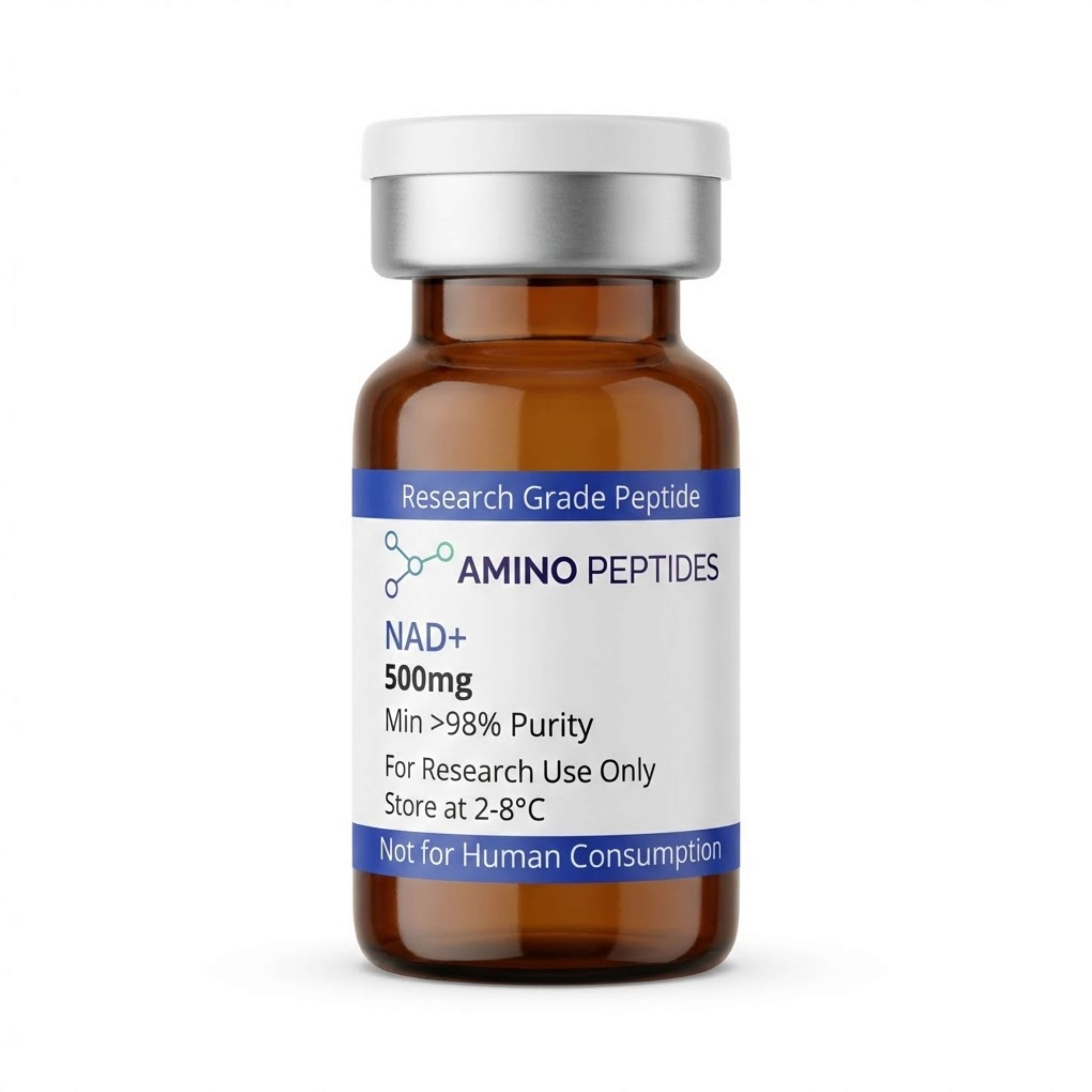
NAD+
From £45
Supplied as a sealed freeze-dried vial (stable 12–24 months at 2°C–8°C in a standard refrigerator).
Shop NAD+ →What Published Studies Observe Across Early And Late Windows
When investigators track NAD+ replenishment in cell cultures and preclinical models, the observed outcomes split clearly into immediate biochemical signalling and slower structural remodelling. Because NAD+ participates in thousands of redox reactions every second, the earliest shifts show up in metabolic readouts long before any physical changes appear in the tissue.
During the first 24 to 72 hours of a typical laboratory protocol, researchers mainly record rapid changes in the intracellular NAD+ to NADH ratio. Within hours of introducing a measured level of NAD+ or its direct precursors to cultured cells, respirometry assays show an uptick in oxygen consumption rates. This early window is defined by immediate enzyme activation. SIRT1 and SIRT3 begin deacetylating target proteins inside the nucleus and mitochondria, which quickly lowers markers of acute oxidative stress. At the same time, PARP enzymes gain the substrate they need to clear a backlog of single-strand DNA breaks, stabilising the genome against further stress.
Structural and tissue-level shifts take much longer to develop in published studies, typically requiring two to eight weeks of consistent protocol ratios in rodent models. Around the two-week mark, researchers begin to observe measurable increases in PGC-1alpha signalling, the regulator responsible for building fresh mitochondria. Rather than just running existing mitochondria harder, the cells start assembling new energy-producing organelles to replace worn-out ones.
By weeks four to eight in preclinical models, these sustained signalling changes translate into visible tissue recovery and improved metabolic stamina. Studies examining muscle and neural tissues during this later phase report healthier mitochondrial cristae structure under electron microscopy, alongside reduced cellular senescence markers. In short, early study windows capture the spark of restored enzyme activity, while multi-week timelines reveal the slower physical work of mitochondrial turnover and long-term genetic maintenance.
Comparison of NAD+ and Related Precursor Molecules in Laboratory Models
| Molecule | Molecular Weight | Primary Cellular Pathway | Membrane Transport | Key Focus in Published Studies |
|---|---|---|---|---|
| NAD+ (Nicotinamide Adenine Dinucleotide) | 663.43 g/mol | Direct cofactor for redox reactions, sirtuins, and PARPs | Requires extracellular breakdown or specific connexin channels | Direct electron transfer, mitochondrial respiration, and genomic repair |
| NMN (Nicotinamide Mononucleotide) | 334.22 g/mol | Direct salvage pathway precursor via NMNAT enzymes | Enters via Slc12a8 transporter or after conversion to NR | Age-related decline models, vascular tissue remodelling, and metabolic signalling |
| NR (Nicotinamide Riboside) | 255.25 g/mol | Converted to NMN inside the cell by NRK1 and NRK2 enzymes | Direct transport across cell membranes via nucleoside transporters | Neuroprotection models, hepatic lipid metabolism, and muscle recovery |
| NAM (Nicotinamide) | 122.12 g/mol | Recycled into NMN via the rate-limiting enzyme NAMPT | Passive diffusion across most mammalian cell membranes | Feedback inhibition of sirtuins at high concentrations and basic salvage recycling |
| NA (Nicotinic Acid) | 123.11 g/mol | Preiss-Handler pathway via NAPRT enzymatic conversion | Active transport via sodium-dependent monocarboxylate transporters | Lipid profile modulation in laboratory models and tissue-specific NAD+ synthesis |
Pairing NAD+ With Complementary Mitochondrial And Repair Peptides
Because NAD+ touches both energy metabolism and structural maintenance, laboratory investigators rarely study it in total isolation. Combining NAD+ with specific research peptides allows scientists to observe how extra coenzyme fuel interacts with targeted receptor signalling. In metabolic protocols, researchers frequently examine MOTS-c and NAD+ side by side to map out mitochondrial efficiency.
MOTS-c is a sixteen-amino-acid peptide encoded directly within mitochondrial DNA. In preclinical models, MOTS-c activates the AMPK pathway, which encourages cells to take up glucose and oxidise fatty acids more efficiently. When a cell's AMPK pathway is switched on by MOTS-c, the demand for electron carriers inside the citric acid cycle rises sharply. Supplying NAD+ alongside MOTS-c in a study ensures that once the peptide signals the cell to burn more fuel, the mitochondria actually have the redox coenzymes required to convert that fuel into ATP without stalling.
Another common laboratory pairing involves SS-31 (Elamipretide). SS-31 binds selectively to cardiolipin on the inner mitochondrial membrane, stabilising the physical curve of the membrane where the electron transport chain sits. While NAD+ drops off the electrons, SS-31 keeps the physical wire intact so fewer electrons leak out as damaging free radicals. Together, they address both the fuel supply and the physical hardware of cellular respiration.
On the structural side, investigators exploring tissue repair research often track how NAD+ availability supports peptides like GHK-Cu or BPC-157. Remodelling collagen matrices and migrating fibroblasts across a scratch assay require enormous amounts of cellular energy and rapid DNA replication. When NAD+ levels are kept steady in the culture medium, sirtuin-driven stress resistance and ATP production remain high, giving structural peptides the energetic backing required to complete complex tissue remodelling tasks.

Figure: NAD+ Research: Tracking Cellular Energy And DNA Repair
Reconstitution calculator
Choose a vial mass and a bacteriostatic reconstitution solution volume. The figure is the resulting concentration.
Concentration: 2.50 mg/mL
Per 0.01mL (10µL): 25.0 mcg
Open the full calculatorBacteriostatic reconstitution solutionWhy 99%+ HPLC Verification And Cold Storage Matter For NAD+
The reliability of any laboratory readout depends heavily on the chemical cleanliness of the starting material in the vial. NAD+ is a relatively large, polar dinucleotide molecule that is notoriously sensitive to heat, light, and moisture. When a batch of NAD+ is poorly purified or exposed to warm shipping conditions, it breaks down into nicotinamide and ADP-ribose before the experiment even begins. High concentrations of free nicotinamide are a major problem in cell assays because nicotinamide actually acts as a feedback inhibitor on sirtuins, meaning a degraded vial can produce the exact opposite signalling result a researcher is trying to measure.
To prevent this, every batch used in quantitative laboratory work should carry a verified High-Performance Liquid Chromatography (HPLC) purity rating of 99% or higher, backed by Mass Spectrometry to confirm the exact molecular weight. Equally important is the removal of trifluoroacetic acid (TFA) salts during the final lyophilisation stages. Residual TFA left over from synthesis can alter the pH of a cell culture medium and prove toxic to delicate mitochondrial membranes, skewing respirometry data.
Proper storage is straightforward but essential for protecting the dinucleotide bond. Lyophilised NAD+ vials belong in a dedicated laboratory refrigerator kept between 2°C and 8°C, shielded from direct light. For multi-month archival storage prior to use, keeping the dry powder at -20°C slows hydrolysis to a crawl. Once a vial is brought to room temperature and mixed with a sterile bacteriostatic reconstitution solution, the solution should be mixed gently by swirling rather than vigorous shaking. Keeping the reconstituted liquid cold at 2°C to 8°C and dividing it into single-use aliquots prevents repeated freeze-thaw cycles from snapping the delicate bond between the nicotinamide and ribose rings.
Frequently Asked Questions
Why is NAD+ research: tracking cellular energy and DNA repair gaining so much scientific attention?
Researchers focus on NAD+ because it acts as a core coenzyme for mitochondrial energy transfer and enzyme activation in all living cells. Published studies show that natural cellular levels decline with age, making it a primary target in laboratory models investigating tissue recovery, cellular longevity, and metabolic signalling pathways.
What timeline do laboratory models typically follow when observing NAD+ and cellular shifts?
In preclinical models, acute shifts in intracellular NAD+ concentrations and ATP output are often recorded within hours of introducing the compound to cell cultures. Broader structural markers, such as sirtuin activation and measurable DNA repair activity, are typically analysed across multi-week study timelines to characterise sustained tissue remodelling.
Which compounds are commonly paired with NAD+ in combined research protocols?
Scientists frequently pair NAD+ with metabolic precursors like NMN or sirtuin activators such as resveratrol to observe synergistic effects in laboratory models. Some protocols also combine it with mitochondrial peptides like MOTS-c or SS-31 to analyse how multiple energy and cellular recovery pathways interact at specific protocol ratios.
How should research-grade NAD+ be stored after mixing to protect its purity?
Lyophilised NAD+ powder should be stored in a freezer at minus 20 degrees Celsius away from light to maintain structural stability. Once mixed with a sterile bacteriostatic reconstitution solution, the liquid must be kept refrigerated between 2 and 8 degrees Celsius and analysed within a few weeks before degradation occurs.
Bibliography
- Covarrubias AJ, Perrone R, Grozio A, Verdin E. NAD+ metabolism and its roles in cellular processes during ageing. Nature Reviews Molecular Cell Biology. 2021;22(2):119-141. View published research
- Rajman L, Chwalek K, Sinclair DA. Potential of NAD-Boosting Molecules: The In Vivo Evidence. Cell Metabolism. 2018;27(3):529-547. View published research
- Yoshino J, Baur JA, Imai SI. NAD+ Intermediates: The Biology and Potential of NMN and NR. Cell Metabolism. 2018;27(3):513-528. View published research
- Verdin E. NAD+ in aging, metabolism, and neurodegeneration. Science. 2015;350(6265):1208-1213. View published research
- Cantó C, Menzies KJ, Auwerx J. NAD+ Metabolism and the Control of Energy Homeostasis: A Balancing Act between Mitochondria and the Nucleus. Cell Metabolism. 2015;22(1):31-53. View published research
⚠️ Research Use Only Disclaimer: All peptides and compounds are sold strictly for in-vitro laboratory research purposes only. Not intended for human or veterinary use, not a dietary supplement, and not approved to diagnose, treat, cure, or prevent any disease or condition. For use by qualified researchers in suitably equipped laboratory environments only. Amino Peptides Ltd operates in full compliance with UK MHRA regulations and applicable EU research chemical guidelines.