
Optimising Culture Conditions to Study the Proliferative Response of Murine Myoblasts to TB-500 Exposure
9th Oct 2026
What You Need to Know About TB-500
- TB-500 is the stocked thymosin beta-4 fragment, searched for actin-pathway and tissue-support research.
- Published cellular work follows actin organisation rather than the fibroblast assays used for BPC-157.
- That is why the two are stacked: the pathways sit next to each other instead of repeating one signal.
- A 99%+ HPLC-verified vial stored at 2°C–8°C is what makes the comparison repeatable.
Growing muscle cells in a laboratory incubator is a delicate balancing act. Murine myoblasts, the precursor cells that eventually form muscle tissue in mice, are notoriously sensitive to their environment. If the nutrient broth is too rich, they multiply rapidly but lose their specific characteristics, becoming useless for targeted research. If the environment lacks key biochemical signals, they stop dividing entirely and simply degrade in the dish. This presents a major hurdle for scientists investigating cellular growth mechanisms. To gather accurate data, researchers must maintain these cells in a state of active division without triggering premature maturation. Recently, attention has turned toward specific synthetic proteins to see if they can stabilise these sensitive cell lines. One major focus of this work involves optimising culture conditions to study the proliferative response of murine myoblasts to TB-500 exposure.
TB-500 is a synthetic version of a specific region found within thymosin beta-4, a naturally occurring protein. In living organisms, thymosin beta-4 plays a critical role in managing actin. Actin is a vital structural protein that acts like the internal scaffolding of a cell. For a cell to divide, move, or change shape, it must constantly break down and rebuild this internal scaffolding. When researchers isolate murine myoblasts in a petri dish, they want to observe how this synthetic fragment interacts with that cellular machinery. The goal is not to force the cells to grow unnaturally, but to map exactly how the peptide influences the natural division cycle under strictly controlled laboratory conditions. By tracking these interactions, scientists can better understand the fundamental rules of cellular structure.
Setting up these experiments requires rigorous preparation. The myoblasts are typically sourced from established laboratory cell lines and thawed carefully. They are placed into plastic culture flasks coated with specific proteins that give the cells a surface to grip. The liquid media covering the cells must be maintained at a precise temperature and pH level, mirroring the internal conditions of a living organism as closely as possible. Once the baseline environment is stable, the researchers prepare the synthetic peptide for introduction.
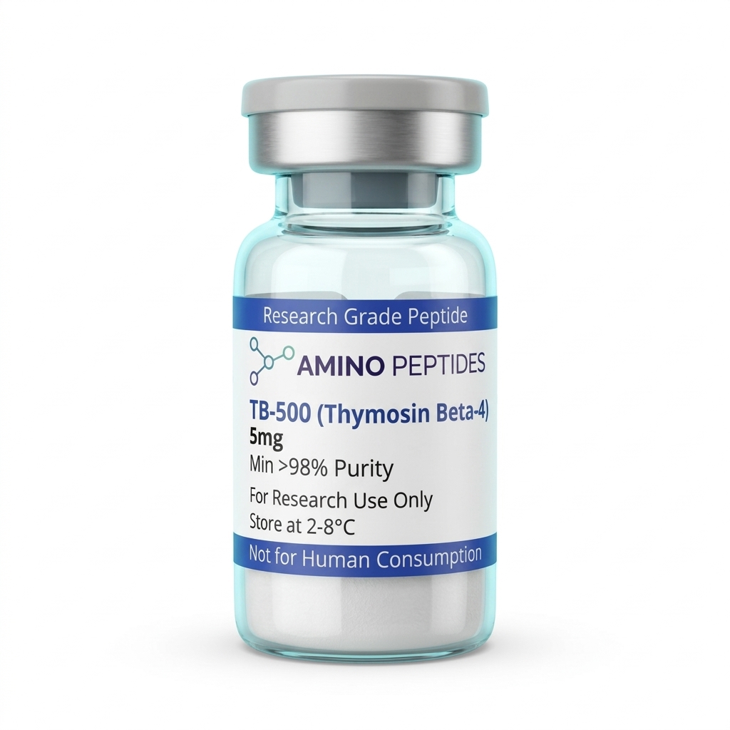
TB-500
From £29
Supplied as a sealed freeze-dried vial (stable 12–24 months at 2°C–8°C in a standard refrigerator).
Shop TB-500 →Once the environment is standardised and the peptide is introduced, scientists monitor the myoblasts over a series of time points, typically 24, 48, and 72 hours. They use automated cell counters and microscopic imaging to track minute changes in the population. The laboratory data shows a clear and repeatable pattern. When exposed to the synthetic fragment, the myoblasts exhibit a higher rate of division compared to control groups kept in standard media. The cells also maintain their intended shape, suggesting the peptide interacts with the internal actin network to support structural integrity during rapid division.
This structural support is largely attributed to a process called actin sequestering. In plain terms, the peptide binds to individual actin molecules floating inside the cell. By holding onto these molecules, the peptide prevents them from linking together into rigid chains before the cell is ready. When the myoblast needs to divide, it requires a flexible internal structure. The peptide essentially acts as a buffer, ensuring there is always a ready supply of free actin building blocks available precisely when the dividing cell needs to reshape its outer membrane.
However, an accelerated division rate in a plastic dish does not equal tissue regeneration in a living organism.
This distinction is critical and often misunderstood. The laboratory environment is an entirely isolated system. It lacks the complex blood supply, immune responses, and hormonal signals present in an actual body. Therefore, the observed proliferation is strictly a biochemical reaction between the peptide and the isolated cellular receptors. Researchers use these findings to understand basic cellular mechanics, not to predict whole-body outcomes. The data simply confirms that the fragment retains its biological activity and can influence the structural proteins of isolated cells. Extrapolating these petri-dish observations to human biology is a fundamental misreading of the scientific method.

Laboratory FAQ: In-Vitro Analysis
What is the standard tb-500 peptide used for in cell cultures?
In laboratory settings, the tb-500 peptide is primarily used as a research reagent to study actin binding and cellular mobility. Scientists apply it to isolated cell lines, such as murine myoblasts, to observe changes in structural integrity and division rates under a microscope. It serves as a tool to map how structural proteins behave during the cell cycle.
Reconstitution calculator
Choose a vial mass and a bacteriostatic reconstitution solution volume. The figure is the resulting concentration.
Concentration: 2.50 mg/mL
Per 0.01mL (10µL): 25.0 mcg
Open the full calculatorBacteriostatic reconstitution solutionHow do researchers design tb 500 protocols for myoblast assays?
Designing tb 500 protocols for in-vitro work requires precise calculation of molar concentrations. Researchers map out specific timeframes for exposure and establish strict control groups that receive only the baseline media. The environment must be tightly regulated, controlling temperature, pH, and nutrient availability to ensure any observed changes in cell count are solely due to the peptide interaction.
What does current tb 500 peptide research indicate about actin binding?
Current tb 500 peptide research indicates that the fragment interacts directly with actin monomers in isolated cells. By binding to these structural proteins, the peptide prevents them from assembling into long chains prematurely. This buffering action appears to help the cell maintain the physical flexibility required to complete division in a petri dish without structural collapse.
What is the standard tb 500 peptide protocol for preparing the reconstitution solvent?
The standard laboratory tb 500 peptide protocol involves dissolving the lyophilised powder in a sterile bacteriostatic reconstitution solution. This solvent prevents bacterial growth in the vial, ensuring the peptide remains stable and uncontaminated during prolonged cellular assays. Proper preparation is essential, as any bacterial presence would rapidly destroy the delicate myoblast culture.
Scientific References
- Philp, D., Nguyen, M., Scheremeta, B., St-Surin, S., Villa, A. M., Orgel, A., Kleinman, H. K., & Elkin, M. (2004). Thymosin beta4 increases hair growth by activation of hair follicle stem cells. The FASEB Journal, 18(2), 385-387. View published research
- Bock-Marquette, I., Saxena, A., White, M. D., DiMaio, J. M., & Srivastava, D. (2004). Thymosin beta4 activates integrin-linked kinase and promotes cardiac cell migration, survival and capillary-like tube formation. Nature, 432(7016), 466-472. View published research
- Goldstein, A. L., Hannappel, E., & Kleinman, H. K. (2005). Thymosin beta4: actin-sequestering protein moonlights to repair injured tissues. Trends in Molecular Medicine, 11(9), 421-429. View published research
- Smart, N., Risebro, C. A., Melville, A. A., Moses, K., Schwartz, R. J., Chien, K. R., & Riley, P. R. (2007). Thymosin beta4 induces adult epicardial progenitor mobilization and neovascularization. Nature, 445(7124), 177-182. View published research
- Sosne, G., Qiu, P., Goldstein, A. L., & Wheater, M. (2010). Biological activities of thymosin beta4 defined by active sites in short peptide sequences. The FASEB Journal, 24(7), 2144-2151. View published research
⚠️ Research Use Only Disclaimer: All peptides and compounds are sold strictly for in-vitro laboratory research purposes only. Not intended for human or veterinary use, not a dietary supplement, and not approved to diagnose, treat, cure, or prevent any disease or condition. For use by qualified researchers in suitably equipped laboratory environments only. Amino Peptides Ltd operates in full compliance with UK MHRA regulations and applicable EU research chemical guidelines.