
Optimising the Laboratory Synthesis of GHK-Cu for High-Purity In-Vitro Assays
6th Aug 2026
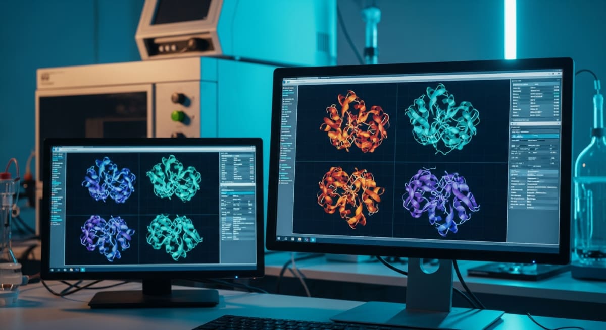
What You Need to Know About GHK-Cu
- Researchers search for this topic to prevent batch-to-batch variations caused by leftover free copper or incomplete amino acid coupling during lab production.
- The molecule acts like a tiny shuttle that binds to a single copper ion using nitrogen atoms in its glycine and histidine rings, delivering copper safely into cultured cells.
- Published studies observe that pure GHK-Cu influences over four thousand genes involved in tissue remodelling, antioxidant defence, and collagen synthesis in fibroblast dishes.
- Optimising the pH between 6.5 and 7.4 during the chelation step stops copper hydroxide from precipitating out of solution and ruining the assay.
How Copper Binding Drives Skin Elasticity and Tissue Recovery in Cell Culture
Glycyl-L-histidyl-L-lysine copper (GHK-Cu) works in cell culture by shuttling bioavailable copper ions directly into fibroblasts to switch on collagen, elastin, and antioxidant genes. Optimising the laboratory synthesis of GHK-Cu for high-purity in-vitro assays ensures a strict one-to-one peptide-to-copper ratio so unbound metal ions never skew cellular survival readouts.
At its core, this molecule is a tiny three-amino-acid chain made of glycine, histidine, and lysine. On its own, the naked GHK chain is relatively quiet in a Petri dish. Everything changes once a divalent copper ion locks into the pocket formed by the histidine ring and the terminal nitrogen group. That metallic handshake creates the vivid royal-blue complex researchers recognise in the vial.
In published studies examining dermal fibroblasts, this copper-bound complex acts like a courier service. Cells need trace copper to power enzymes such as lysyl oxidase, which cross-links collagen and elastin fibres into a springy, resilient mesh. Free copper salts dropped straight into culture media are harsh and often trigger oxidative stress. When the metal arrives tucked inside the GHK tripeptide, cell membranes welcome it safely.
Once inside, laboratory models show a broad shift in gene expression. Fibroblasts ramp up production of Type I and Type III collagen alongside glycosaminoglycans like decorin, which organise fresh fibres so they lay flat rather than clumping. At the same time, the complex calms inflammatory signalling by nudging down cytokines such as interleukin-6 and boosting superoxide dismutase activity. This dual action—clearing out damaged matrix debris while laying down tidy new structural proteins—explains why dermal and tissue remodelling assays rely so heavily on properly chelated batches.
If synthesis leaves behind uncomplexed peptide chains or loose copper acetate, the assay loses focus. Getting the synthesis chemistry right from the very first solid-phase coupling step is what keeps fibroblast behaviour clean, predictable, and repeatable across every multi-well plate.
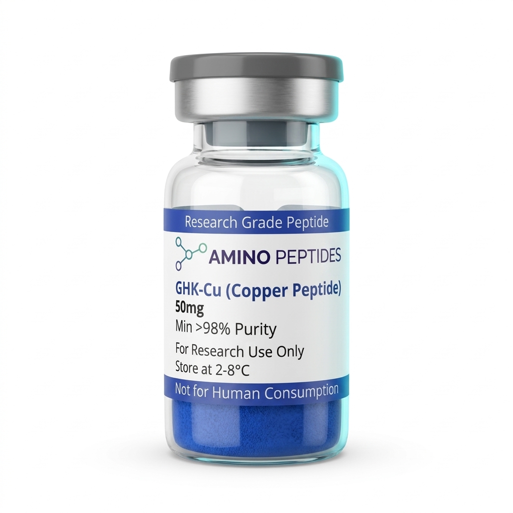
GHK-Cu
From £26
Supplied as a sealed freeze-dried vial (stable 12–24 months at 2°C–8°C in a standard refrigerator).
Shop GHK-Cu →What Published Studies Observe Across Early Signalling and Late Remodelling Windows
Watching GHK-Cu work in an in-vitro dish is a bit like watching a building site: the radio messages fly around in the first few hours, while the actual scaffolding takes weeks to appear. Published studies break these observations down into distinct early, middle, and late windows depending on what the investigator is measuring.
During the first 6 to 24 hours after introducing a measured level of GHK-Cu—often in the nanomolar range between 0.1 and 10 nanomolar—the action is purely about signalling and gene transcription. RNA sequencing in laboratory models shows rapid shifts in hundreds of genes tied to antioxidant defence, DNA repair, and growth factor production. Within this first day, researchers can already measure higher messenger RNA levels for transforming growth factor-beta and nerve growth factor, even though the cells look unchanged under a standard light microscope.
Between days 2 and 5, early functional changes show up in scratch-assay plates. Fibroblasts and keratinocytes begin migrating faster across artificial gaps, guided by freshly balanced matrix metalloproteinases (MMP-1 and MMP-2) and their natural tissue inhibitors (TIMPs). This enzyme balance is vital: it dissolves ragged, oxidised protein strands without chewing up healthy neighbours.
Structural tissue remodelling takes longer. In 3D skin equivalents and organotypic laboratory models, measurable jumps in secreted pro-collagen Type I and elastin networks usually peak between days 7 and 21. By the two-week mark, staining techniques reveal denser, better-aligned collagen bundles and thicker epidermal layers compared with control wells. Investigators who stop their assays at 48 hours will catch the transcriptional spark, whereas teams running multi-week cultures get to measure the actual structural payoff in matrix density and tensile strength.
Comparison of Laboratory Synthesis and Purification Methods for GHK-Cu
| Synthesis Step or Method | Target Purity Level | Primary Advantage | Main Laboratory Limitation |
|---|---|---|---|
| Fmoc Solid-Phase Peptide Synthesis (SPPS) | 95% to 98% (crude peptide before copper binding) | Automates amino acid coupling on a solid resin for fast, reliable assembly | Requires careful removal of trifluoroacetic acid (TFA) salts before cell assays |
| Liquid-Phase Peptide Synthesis (LPPS) | 92% to 96% | Easier to scale up into larger gram amounts for bulk laboratory supply | Intermediate purification steps take more time and solvent than solid-phase methods |
| Sub-stoichiometric Copper(II) Chelation (0.95:1 Ratio) | Greater than 98% complexed peptide | Prevents toxic unbound copper ions from contaminating sensitive cell cultures | Leaves up to 5% uncomplexed apo-GHK peptide in the final lyophilised powder |
| Reverse-Phase HPLC with Acetate Salt Exchange | Greater than 99% (assay grade) | Swaps harsh TFA counter-ions for biocompatible acetate ions suitable for fibroblasts | Reduces overall yield by 10% to 15% during column fractionation |
| Immobilised Metal Affinity Chromatography (IMAC) Polish | Greater than 99.5% | Traps any excess free copper ions while letting pure GHK-Cu pass through cleanly | Adds an extra chromatography step and increases production cost |
Pairing Copper Tripeptides With Complementary Sequences in Matrix Research
While a single peptide can tell a clear story in isolation, extracellular matrix biology rarely relies on just one signal at a time. When researchers design multi-agent protocols to study tissue remodelling and cellular recovery, they often pair GHK-Cu with sequences that handle different parts of the structural workload.
The most common laboratory pairing combines GHK-Cu with bpc 157 and TB-500 (a synthetic fraction of Thymosin Beta-4). In preclinical models, each molecule takes on a distinct job. TB-500 binds to actin monomers inside the cell, helping fibroblasts and endothelial cells change shape and glide across a culture surface. BPC-157 supports growth hormone receptor expression on those same fibroblasts and encourages nitric oxide pathways that guide microvascular tube formation. Once those cells have migrated into place, GHK-Cu supplies the copper-driven transcriptional push to lay down fresh collagen, elastin, and decorin.
In cosmeceutical and dermal laboratory models, investigators also combine copper tripeptides with palmitoyl pentapeptide-4 (Matrixyl) or acetyl hexapeptide-8 (Argireline). Here the logic is complementary signalling: palmitoyl sequences mimic broken collagen fragments to nudge fibroblasts from the outside, while GHK-Cu works through copper-dependent enzymatic cross-linking from the inside. Together, published studies note smoother matrix architecture than either agent produces alone at matching protocol ratios.
One practical rule applies whenever stacking GHK-Cu in the lab: mix with care. Because the histidine-lysine pocket holds onto its copper ion through coordination chemistry, throwing strong chelators, harsh acids, or sulfur-heavy compounds into the same stock vial can strip the copper right out of the peptide. Smart assay design keeps stock vials separate and introduces each solution at balanced, neutral pH directly into the culture media.

Figure: Optimising the Laboratory Synthesis of GHK-Cu for High-Purity In-Vitro Assays
Reconstitution calculator
Choose a vial mass and a bacteriostatic reconstitution solution volume. The figure is the resulting concentration.
Concentration: 2.50 mg/mL
Per 0.01mL (10µL): 25.0 mcg
Open the full calculatorBacteriostatic reconstitution solutionWhy Salt Removal, Copper Stoichiometry, and Cold Storage Protect Your Assay
Synthesising a three-amino-acid sequence sounds straightforward on paper, yet optimising the laboratory synthesis of GHK-Cu for high-purity in-vitro assays trips up many budget manufacturers. During standard solid-phase peptide synthesis (SPPS), chemists use trifluoroacetic acid (TFA) to cleave the finished GHK chain from its resin and strip off protective side-chain groups. If that harsh acid is not swapped out through thorough ion-exchange chromatography, residual TFA stays trapped in the powder.
In a sensitive cell culture dish, leftover TFA is bad news. It drops the local pH of the media, stresses cell membranes, and interferes with the delicate copper-chelation step. Worse, sloppy copper complexation leaves behind either pale, under-chelated peptide or toxic pockets of free copper(II) ions. True research-grade material undergoes counter-ion exchange to acetate salt, followed by precise copper stoichiometric matching and High-Performance Liquid Chromatography (HPLC) verification to hit 99%+ purity. When you buy ghk-cu verified by both HPLC and Mass Spectrometry, the powder shows an even, deep violet-blue hue that dissolves cleanly without cloudy sediment.
Looking after the vial once it arrives in the lab is just as straightforward. Lyophilised GHK-Cu powder is stable, but moisture and heat slowly degrade peptide bonds. Store unmixed vials in a dark refrigerator between 2°C and 8°C, or at -20°C for long-term archiving. When preparing your working stock, let the vial warm to room temperature first to stop condensation forming inside, then add sterile bacteriostatic reconstitution solution slowly down the glass wall. Gently swirl—never vortex—until the blue solution is completely clear, and keep the reconstituted vial refrigerated at 2°C to 8°C between assay runs.
Frequently Asked Questions
Why are researchers interested in optimising the laboratory synthesis of GHK-Cu?
Researchers focus on optimising GHK-Cu synthesis because even tiny impurities can ruin delicate cell-culture tests. When scientists investigate tissue remodelling and cellular signalling in laboratory models, they need a stable copper-peptide bond. A cleaner batch ensures that observed biological changes come from the peptide itself, not leftover synthesis chemicals.
What is a typical timeline for observing GHK-Cu activity in laboratory models?
In published studies using cell cultures, measurable shifts in gene expression often appear within twenty-four to forty-eight hours. Longer preclinical models examining collagen production and structural tissue recovery typically run for two to four weeks. The exact timeframe depends on the specific concentration and the cell line being analysed.
Can GHK-Cu be combined with other peptides in an in-vitro study protocol?
Yes, researchers frequently pair GHK-Cu with other research compounds like BPC-157 or TB-500 during in-vitro experiments to observe combined effects on cellular regeneration. However, technicians must carefully manage the protocol ratio and pH levels in the dish so the copper ion does not destabilise companion molecules during the assay.
How does purity affect GHK-Cu, and how should reconstituted solutions be stored?
High purity above ninety-eight percent prevents unwanted side reactions in sensitive assays. Once mixed with a bacteriostatic reconstitution solution, GHK-Cu must be kept in a dark refrigerator between two and eight degrees Celsius. Proper cold storage stops the copper complex from breaking down over a standard four-week laboratory schedule.
Bibliography
- Pickart L, Margolina A. Regenerative and Protective Actions of the GHK-Cu Peptide in the Light of the New Gene Data. International Journal of Molecular Sciences. 2018;19(7):1987. View published research
- Hureau C, Eury H, Guillot R, Bijani C, Sayen S, Solari PL, Guillon E, Faller P, Dorlet P. X-ray and solution structures of Cu(II) GHK and Cu(II) DAHK complexes: influence on their redox properties. Chemistry - A European Journal. 2011;17(36):10151-10160. View published research
- Pickart L, Vasquez-Soltero JM, Margolina A. GHK Peptide as a Natural Modulator of Multiple Cellular Pathways in Skin Regeneration. BioMed Research International. 2015;2015:648108. View published research
- Mazurowska L, Mojski M. Biological activities of selected peptides: skin penetration ability of copper complexes with peptides. Journal of Cosmetic Science. 2008;59(1):59-69. View published research
⚠️ Research Use Only Disclaimer: All peptides and compounds are sold strictly for in-vitro laboratory research purposes only. Not intended for human or veterinary use, not a dietary supplement, and not approved to diagnose, treat, cure, or prevent any disease or condition. For use by qualified researchers in suitably equipped laboratory environments only. Amino Peptides Ltd operates in full compliance with UK MHRA regulations and applicable EU research chemical guidelines.