
Quantifying Purity: How HPLC and MS Validation Protect Your Research Integrity
22nd Sep 2026
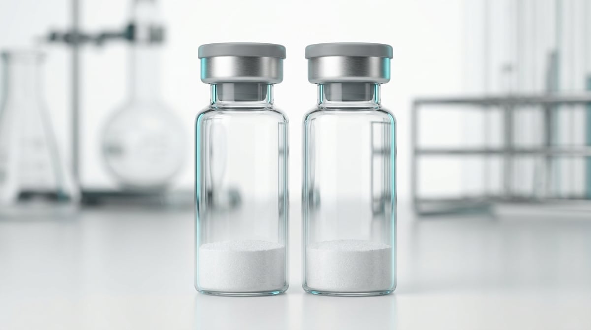
What You Need to Know About Quantifying Purity: How HPLC and MS Validation Protect Your Research Integrity
- Researchers search for this topic to learn how to read Certificates of Analysis and prevent contaminated reagents from ruining months of laboratory work.
- High-Performance Liquid Chromatography pushes dissolved molecules through a tightly packed column to separate the target peptide from leftover synthesis byproducts and measure its exact percentage purity.
- Mass Spectrometry weighs the separated molecules at the atomic level to confirm that the amino acid sequence matches the expected molecular weight.
- Published studies consistently demonstrate that unverified peptides often contain truncated sequences and residual synthesis acids that distort cellular signalling assays and reduce reproducibility.
How HPLC and Mass Spectrometry Guard Cellular Energy and Recovery Data
High-Performance Liquid Chromatography (HPLC) measures exact chemical purity by separating a sample into its individual components, whilst Mass Spectrometry (MS) weighs the molecules to prove the exact amino acid sequence is present. Together, these two tests ensure that laboratory observations in cellular energy, skin elasticity, or tissue recovery come from the target peptide rather than hidden synthesis leftovers.
When chemists build a peptide chain in a laboratory, they link amino acids together one step at a time. Sometimes a link fails to attach, leaving behind a shorter, broken chain called a deletion sequence. Even a small fraction of these broken chains can ruin an experiment. If a team is studying how a specific sequence supports cellular energy in isolated mitochondria or promotes collagen organisation for skin elasticity, a broken fragment might block the target receptor without switching it on. That silent blockage makes an active compound look weak or useless.
HPLC solves this problem by pushing the dissolved peptide through a tightly packed column under high pressure. Different molecules travel through this column at different speeds depending on how strongly they cling to the internal packing material. A detector at the exit shines ultraviolet light through the liquid and draws a graph of peaks. The main peak represents the target peptide, and the area inside that peak tells analysts the exact percentage of purity. However, HPLC alone cannot prove identity, because two completely different molecules might happen to travel at the exact same speed.
That is why Mass Spectrometry is the essential second half of the test. MS gives the molecules an electrical charge, accelerates them through a vacuum, and measures their mass-to-charge ratio down to a fraction of a Dalton. Every amino acid has a known atomic weight, so the complete peptide has a strict mathematical fingerprint. When the MS readout matches the theoretical molecular weight and the HPLC graph shows a clean single peak, researchers know their recovery or focus assay is starting with verified material.
This page is not tied to one stocked vial. Start with the bundles hub, BPC-157, or GHK-Cu — each is in stock for next-day UK dispatch.
Browse Full UK Peptide Catalogue →What Degraded Samples Do to Early Signalling and Late Tissue Remodelling
In published studies, the cleanliness of a reagent shapes every stage of an observation timeline, from the first few minutes of receptor binding to weeks of structural tissue remodelling. During the early signalling window, which usually spans the first fifteen minutes to six hours in cell culture, peptides bind to surface receptors and trigger fast internal messengers like cyclic AMP or calcium waves. If a vial has not been properly verified by HPLC and MS, truncated fragments can compete for those exact same docking sites. Instead of seeing a sharp, clean spike in early signalling, the laboratory readout looks flat or erratic within the first hour.
As a study moves into the intermediate window of twenty-four to seventy-two hours, researchers typically look for changes in gene expression and protein production. At this stage, chemical leftovers from manufacturing cause a different set of problems. Unremoved protecting groups or oxidised side chains can stress the cells, switching on defensive inflammatory pathways that have nothing to do with the actual study. In preclinical models examining sleep architecture or neural focus signals, these background contaminants create confusing noise that masks the gentle, natural rhythm of the target molecule.
The damage becomes even clearer during long-term observations lasting two to six weeks, where the focus shifts to visible structural changes like collagen matrix assembly, blood vessel sprouting, or muscle fibre recovery. Structural remodelling requires steady, predictable biochemical signals over many days. When a batch contains five or ten percent unknown impurities, those mystery compounds build up in the culture medium over repeated study amounts. By week three, fibroblast cells that should be laying down neat, elastic structural fibres may slow down or show signs of toxicity. Comparing a verified 99 percent pure batch against an unverified 92 percent batch over a four-week timeline consistently shows that clean starting material is the only way to track genuine structural recovery from start to finish.
Comparison of Analytical Validation Methods in Peptide Research
| Analytical Technique | Primary Measurement | What It Detects | Limitation If Used Alone |
|---|---|---|---|
| RP-HPLC (Reversed-Phase HPLC) | Relative purity percentage by ultraviolet absorbance | Organic peptide impurities, deletion sequences, and degraded fragments | Cannot confirm the exact molecular identity or detect invisible inorganic salts |
| Mass Spectrometry (ESI-MS / MALDI-TOF) | Mass-to-charge ratio (m/z) and exact molecular weight | Target sequence identity, oxidation states, and specific synthesis modifications | Does not provide an accurate percentage of overall sample purity on its own |
| Ion Chromatography (IC) / Counter-Ion Analysis | Concentration of bound counter-ions and residual salts | Trifluoroacetic acid (TFA), acetate, and chloride levels left over from purification | Provides zero information about the peptide sequence or organic purity |
| Karl Fischer Titration | Residual moisture content by weight | Water molecules trapped inside the freeze-dried peptide powder | Only measures water content and cannot detect peptide degradation |
| Amino Acid Analysis (AAA) | Net peptide content and amino acid ratios | Exact amount of active peptide versus total powder weight including salts and water | Destroys the sample during acid hydrolysis and cannot spot sequence order errors |
Why Multi-Peptide Stacks Demand Twin Analytical Verification
Combining two or more complementary compounds in a single experimental protocol is a popular way to study how different biological pathways cooperate, yet multi-peptide stacks multiply the risk of chemical cross-reactions if either vial lacks proper analytical verification. When researchers pair regenerative sequences or metabolic fragments together, they rely on each molecule doing its specific job without interfering with its partner. If one vial carries hidden synthesis by-products, those stray chemicals can react with the second peptide in solution, ruining both compounds before they ever touch a cell culture.
Consider a metabolic research design that pairs a growth hormone fragment like the aod9064 peptide with a mitochondrial support sequence to observe fat breakdown alongside cellular energy output. The lipolytic fragment relies on a delicate disulfide bridge between two cysteine amino acids to hold its folded shape. If the companion peptide in the stack was poorly purified and still contains trace oxidising agents or acidic scavengers from the factory, mixing or running them side by side can snap that disulfide bridge. MS validation catches oxidised variants early, showing a tell-tale mass shift of sixteen Daltons, which warns the analyst that the molecule has picked up an extra oxygen atom.
The same strict requirement applies when investigators combine immune-modulating and barrier-support sequences such as KPV and LL-37 to study how epithelial cells calm inflammation whilst defending against microbial stress. KPV is a tiny three-amino-acid sequence, whereas LL-37 is a much larger thirty-seven-amino-acid chain with a strong positive charge. Because LL-37 is longer and harder to assemble, unverified batches frequently contain thirty-five- or thirty-six-amino-acid deletion sequences that look nearly identical on a basic test. Only high-resolution HPLC paired with electrospray ionisation MS can separate and weigh those close cousins, ensuring that a dual-pathway synergy study reflects true biological teamwork rather than a clash of broken fragments.

Figure: Quantifying Purity: How HPLC and MS Validation Protect Your Research Integrity
Reading the Certificate: 99%+ Benchmarks, Salt Removal, and Cold Storage
Checking a batch-specific Certificate of Analysis (CoA) before starting any laboratory protocol protects months of hard work from preventable chemical failure. A trustworthy CoA always displays two actual instrument printouts rather than just a typed table of numbers: the HPLC chromatogram showing a purity peak of 99 percent or higher, and the MS spectrum showing the exact mass ions. When the HPLC trace shows a sharp, narrow peak with a flat baseline and no messy shoulders, the vial contains a uniform population of molecules ready for precise measured levels.
Beyond the peptide itself, smart vial quality control also looks at counter-ions, especially Trifluoroacetic Acid (TFA). TFA is a strong acid used during solid-phase manufacturing to cleave the finished peptide off its resin bead and keep it dissolved during HPLC purification. If left behind in high amounts, residual TFA forms a salt with the peptide that can lower the pH of a cell culture and prove toxic to delicate mammalian cells, skewing proliferation and recovery assays. Premium research vials undergo an extra ion-exchange step or thorough lyophilisation to strip away excess TFA, replacing it with benign acetate salts or reducing the acid footprint to harmless trace levels so cellular assays stay healthy.
Once a verified 99 percent pure lyophilised powder arrives in the laboratory, keeping that analytical score intact comes down to simple temperature discipline. Dry peptide cake is stable for transport, but long-term storage requires a dedicated refrigerator kept between 2°C and 8°C, away from bright light and moisture. Before opening a cold vial, let it warm up to room temperature inside a dry desiccator for twenty minutes so room humidity does not condense onto the powder. When preparing a liquid stock using bacteriostatic reconstitution solution, gently trickle the liquid down the inside glass wall and swirl rather than shaking, preserving the exact folding and purity that the HPLC and MS machines confirmed at the laboratory.
Frequently Asked Questions
Why do researchers search for HPLC and MS validation data before starting a study?
Researchers verify HPLC and MS data to ensure their compounds are authentic and free from hidden contaminants. Without independent verification, leftover synthesis chemicals can skew cellular signalling in laboratory models. Checking certificates of analysis protects the experiment, saves money, and guarantees that published studies reflect accurate, repeatable scientific observations.
How long does analytical validation take before a laboratory study timeline can begin?
Third-party HPLC and mass spectrometry testing typically takes five to ten working days before a laboratory timeline can safely begin. Analytical technicians dissolve a small sample, run it through separation columns, and characterise the molecular weight. Waiting for this verified report prevents wasted weeks on flawed preclinical models.
Can researchers combine multiple peptides in one assay, and how is purity verified?
Researchers frequently combine multiple compounds in a single assay, but each must be validated individually before mixing. When stacking reagents, HPLC confirms that neither compound contains synthesis byproducts that might cross-react. Analyzing each vial separately ensures the combined protocol ratio delivers clean, reliable data during tissue remodelling observations.
How does fridge storage affect verified peptide purity after adding bacteriostatic reconstitution solution?
Once mixed with bacteriostatic reconstitution solution, a peptide's verified purity slowly declines even under proper fridge storage at four degrees Celsius. Liquid mixtures are vulnerable to oxidation and chain breakdown over several weeks. To optimise experimental integrity, laboratories keep lyophilised powders frozen and only mix small study amounts as needed.
Bibliography
- D'Hondt M, Bracke N, Taevernier L, Gevaert B, Verbeke F, Wynendaele E, De Spiegeleer B. Related impurities in peptide analytical profiles: a review. Journal of Pharmaceutical and Biomedical Analysis. 2014;101:2-30. View published research
- Cornish J, Callon KE, Lin CQ, Xiao CL, Mulvey TB, Cooper GJ, Reid IR. Trifluoroacetate, a contaminant in purified proteins, inhibits proliferation of osteoblasts and chondrocytes. American Journal of Physiology. 1999;277(5):E779-E783. View published research
- Mant CT, Chen Y, Yan Z, Popa TV, Kovacs JM, Mills JB, Tripet BP, Hodges RS. HPLC analysis and purification of peptides. Methods in Molecular Biology. 2007;386:3-55. View published research
- Boesl U. Time-of-flight mass spectrometry: Introduction to the basics. Mass Spectrometry Reviews. 2017;36(1):86-109. View published research
- Begley CG, Ioannidis JP. Reproducibility in science: improving the standard for basic and preclinical research. Circulation Research. 2015;116(1):116-126. View published research
See our UK peptide research guides & COA briefings first in Google Search.
Add Amino Peptides UK as a Preferred Source on Google⚠️ Research Use Only Disclaimer: All peptides and compounds are sold strictly for in-vitro laboratory research purposes only. Not intended for human or veterinary use, not a dietary supplement, and not approved to diagnose, treat, cure, or prevent any disease or condition. For use by qualified researchers in suitably equipped laboratory environments only. Amino Peptides Ltd operates in full compliance with UK MHRA regulations and applicable EU research chemical guidelines.