
Scientific Analysis of MOTS-c: Mitochondrial-Derived Peptide Research
26th Jun 2026

What You Need to Know About MOTS-C
- Curious readers often search for MOTS-c to understand how mitochondria send chemical signals that mimic the cellular benefits of physical exercise.
- It works by pausing the folate cycle inside the cell, which causes a natural build-up of an intermediate molecule that switches on the master energy sensor AMPK.
- Published studies in preclinical models show that this peptide moves into the cell nucleus during metabolic stress to help regulate genes involved in glucose uptake and antioxidant defence.
- Laboratory researchers have observed improved insulin sensitivity, better cold tolerance, and stronger physical endurance in aged mice given measured levels of MOTS-c.
How MOTS-c Drives Cellular Energy Through AMPK Signalling
MOTS-c is a sixteen-amino-acid peptide encoded directly within mitochondrial DNA that regulates cellular energy by activating the AMPK pathway. In laboratory models, this signalling action helps cells process glucose efficiently and adapt to metabolic stress.
Most peptides studied in modern laboratories are encoded in the cell's nucleus. MOTS-c breaks that rule completely. Discovered in 2015, its genetic instructions live inside the 12S rRNA region of the mitochondria, the tiny power plants inside almost every living cell. Because of this unusual origin, scientists classify it as a mitochondrial-derived peptide. When a cell experiences energy stress, such as low glucose availability or intense physical demand in preclinical models, MOTS-c travels out of the mitochondria and into the main cell body, and even into the nucleus itself, to help organise a clear metabolic response.
Once active, MOTS-c targets the folate-methionine cycle. Specifically, it slows down a step in the folate cycle, which causes a natural compound called AICAR to build up inside the cell. Researchers recognise AICAR as a direct activator of AMP-activated protein kinase, better known as AMPK. You can think of AMPK as the cell's fuel gauge. When energy reserves drop, AMPK flips on to restore balance. It encourages the cell to pull more glucose out of the surrounding fluid and burn fatty acids for fuel instead of storing them.
In published studies examining skeletal muscle cultures, this AMPK activation prompts glucose transporter type 4 (GLUT4) to move to the cell surface. That movement allows glucose to enter the muscle cell without relying solely on heavy insulin signalling. At the same time, when MOTS-c enters the cell nucleus during periods of metabolic stress, it binds to DNA regions controlled by the transcription factor NRF2. This binding turns on protective genes that help the cell survive oxidative strain and maintain steady energy output.
Rather than acting as a stimulant that forces a cell to work harder, MOTS-c functions in laboratory models as an efficiency coordinator. It helps tissues make better use of the fuel already sitting in front of them, which explains why metabolic researchers study it so closely for cellular energy, endurance adaptation, and age-related metabolic decline.
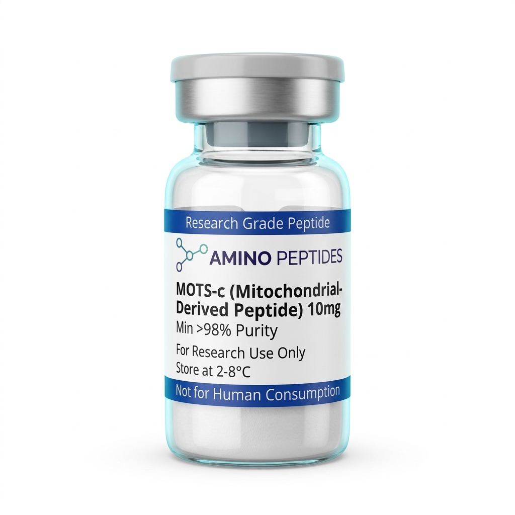
MOTS-C
From £30
Supplied as a sealed freeze-dried vial (stable 12–24 months at 2°C–8°C in a standard refrigerator).
Shop MOTS-C →What Published Studies Observe Across Early and Late Timelines
When researchers design a scientific analysis of MOTS-c: mitochondrial-derived peptide research, they separate their observations into immediate biochemical signalling and longer-term structural adaptation. Because MOTS-c works by adjusting how cells handle fuel and stress genes, the timeline in preclinical models moves in distinct stages rather than all at once.
During the first 24 to 72 hours of exposure in cell culture and rodent studies, the changes are purely chemical and happen inside the cell's energy pathways. Within hours of introducing a measured level of MOTS-c, laboratory assays detect a sharp rise in endogenous AICAR levels and immediate phosphorylation of AMPK. During this early window, cultured muscle cells show higher glucose clearance from their growth medium and a rapid shift toward fatty acid oxidation. No physical tissue remodelling has occurred yet; the cells are simply flipping their metabolic switches and clearing out excess glucose more briskly.
Between week one and week three of continuous observation in animal models, those early chemical signals begin to translate into functional stamina and metabolic steadiness. Published studies tracking mice on high-fat diets or treadmill endurance tests note measurable shifts in how the whole organism handles fuel. Blood glucose curves flatten out, insulin sensitivity markers improve in skeletal muscle, and test subjects run noticeably longer before reaching exhaustion. At this midpoint, gene expression arrays also show sustained activity in NRF2-linked antioxidant pathways, meaning the tissues are actively defending themselves against the oxidative byproducts of heavy energy production.
From week four through week eight and beyond, preclinical models reveal physical tissue remodelling and structural adaptation. In aged rodent models, longer protocols are associated with better maintenance of lean muscle mass, reduced fat accumulation around internal organs, and higher mitochondrial density inside muscle fibres. High-resolution imaging of these muscle tissues shows healthier mitochondrial structure and improved respiratory capacity compared to untreated control groups. In short, the early hours are all about AMPK activation and fuel uptake, the middle weeks bring measurable endurance and glucose handling changes in the lab, and the later weeks show physical upkeep of the mitochondrial network itself.
Comparison of Mitochondrial-Derived Peptides in Laboratory Research
| Peptide Name | Mitochondrial Gene Origin | Amino Acid Length | Primary Signalling Pathway | Key Observation in Preclinical Models |
|---|---|---|---|---|
| MOTS-c | 12S rRNA (MT-RNR1) | 16 amino acids | Folate-AICAR-AMPK pathway and NRF2 nuclear translocation | Regulates skeletal muscle glucose metabolism and enhances physical capacity in aged rodent models |
| Humanin | 16S rRNA (MT-RNR2) | 24 amino acids | IGFBP-3 binding, BAX inhibition, and STAT3 signalling | Protects cells against oxidative stress and suppresses programmed cell death in neural and vascular tissue cultures |
| SHLP2 | 16S rRNA (MT-RNR2) | 26 amino acids | ERK and STAT3 activation with mitochondrial oxygen consumption support | Improves cellular energy output and supports metabolic homeostasis in age-related laboratory models |
| SHLP6 | 16S rRNA (MT-RNR2) | 20 amino acids | Apoptotic cascade modulation | Increases programmed cell death in specific abnormal cell lines while sparing healthy control cells in vitro |
Pairing MOTS-c with Complementary Mitochondrial Research Compounds
Because cellular energy production involves dozens of overlapping steps, laboratory investigators rarely look at a single molecule in isolation. In preclinical models, combining MOTS-c with other mitochondrial and metabolic compounds allows researchers to observe how different stages of the energy cycle support one another without overloading a single receptor pathway.
One of the most frequent combinations in metabolic research pairs MOTS-c and NAD+ in the same experimental protocol. The logic behind this pairing is straightforward. While MOTS-c activates AMPK to bring glucose and fatty acids into the cell's furnace, nicotinamide adenine dinucleotide (NAD+) acts as the essential coenzyme that carries electrons through the Krebs cycle and the electron transport chain. Furthermore, AMPK activation naturally nudges the cell to recycle more NAD+, which in turn fuels sirtuin enzymes like SIRT1 and SIRT3 that keep mitochondria healthy. Observing both compounds together lets researchers study fuel delivery and electron transport at the exact same time.
Another natural companion in mitochondrial studies is SS-31, also known as Elamipretide. While MOTS-c operates as a signalling peptide that talks to the folate cycle and the cell nucleus, SS-31 has a purely structural job inside the inner mitochondrial membrane. SS-31 binds directly to cardiolipin, a special phospholipid that holds the electron transport chain complexes in place. By stabilising cardiolipin, SS-31 reduces electron leak and oxidative damage right at the hardware level. Pairing the structural membrane support of SS-31 with the AMPK signalling of MOTS-c gives laboratory models both software instructions and hardware protection.
Researchers also frequently evaluate MOTS-c alongside Humanin, another peptide encoded in the mitochondrial 16S rRNA region. Where MOTS-c focuses on glucose metabolism and muscle endurance, Humanin is studied primarily for cytoprotection and shielding cells from stress-induced apoptosis. For laboratories investigating multi-compound mitochondrial protocols, a mitoprime peptide setup often serves as a convenient way to study how these distinct mitochondrial pathways interact across shared cellular environments.
Reconstitution calculator
Choose a vial mass and a bacteriostatic reconstitution solution volume. The figure is the resulting concentration.
Concentration: 2.50 mg/mL
Per 0.01mL (10µL): 25.0 mcg
Open the full calculatorBacteriostatic reconstitution solutionWhy 99%+ HPLC Purity and Cold Storage Protect Your Vial
A sixteen-amino-acid sequence like MOTS-c relies on its exact three-dimensional fold to interact with the folate cycle and bind to nuclear DNA regions. If a batch contains truncated peptide chains, leftover synthesis solvents, or moisture damage, the experimental data will be skewed before the study even begins. That is why checking the analytical paperwork of the vial itself is the smartest habit any laboratory buyer can build.
Every reliable research vial should be backed by High-Performance Liquid Chromatography (HPLC) and Mass Spectrometry (MS) reports confirming 99%+ purity. During solid-phase peptide synthesis, amino acids are linked together one by one. Occasionally, an amino acid fails to attach, leaving behind a fifteen-amino-acid fragment that looks nearly identical to MOTS-c but lacks its biological signalling ability. HPLC separates and measures the exact percentage of the full, intact peptide versus those incomplete fragments, while Mass Spectrometry confirms the molecular weight matches the true theoretical mass of MOTS-c (2174.6 Daltons).
Equally important is the removal of trifluoroacetic acid (TFA). TFA is a strong acid used during manufacturing to cleave the finished peptide from its resin bead. If a supplier skips the ion-exchange polishing step, residual TFA remains bound to the peptide powder as a salt. In cell culture models, excess TFA lowers the pH of the medium, stresses living cells, and creates false toxicity readings that have nothing to do with MOTS-c itself. Premium research preparations swap out harsh TFA ions for benign acetate salts, keeping the cellular environment clean and neutral.
Once the lyophilised (freeze-dried) powder arrives, protecting the vial from heat and light keeps the amino acid chain intact. Unreconstituted vials should be stored in a freezer at -20°C for long-term archiving. After the powder is gently mixed with a bacteriostatic reconstitution solution—letting the liquid slide down the glass wall rather than shaking the vial—the solution must be kept in a refrigerator between 2°C and 8°C. At that cool temperature, the peptide bonds resist hydrolysis and oxidation, preserving clean, repeatable signalling across the entire study window.
Frequently Asked Questions
Why are researchers investigating MOTS-c so closely?
Researchers study MOTS-c because it is a unique peptide encoded directly inside mitochondrial DNA that helps regulate cellular energy and metabolic homeostasis. Published studies show it activates the AMPK signalling pathway in laboratory models. This helps scientists analyse how cells adapt to metabolic stress, exercise demands, and age-related cellular decline.
What is the typical timeline observed in MOTS-c laboratory studies?
In preclinical models, acute cellular signalling shifts often appear within hours of exposure, while measurable metabolic adaptations typically take two to eight weeks. Researchers tracking physical endurance and glucose regulation in rodent models usually observe steady changes across a four-week study window, depending on the exact protocol ratio.
Can MOTS-c be combined with other mitochondrial peptides in research?
Yes, laboratory protocols frequently pair MOTS-c with other mitochondrial-derived peptides like Humanin or SS-31 to analyse complementary cellular pathways. While MOTS-c focuses on AMPK signalling and glucose metabolism, partner compounds often target oxidative stress. Combining them allows scientists to observe broader cellular recovery and energy optimisation in preclinical models.
How should MOTS-c be stored to maintain high research purity?
Lyophilised MOTS-c powder should be stored in a freezer at minus 20 degrees Celsius away from bright light to preserve its structural purity. Once mixed with a bacteriostatic reconstitution solution for laboratory analysis, the liquid must be kept refrigerated between 2 and 8 degrees Celsius and used within three weeks.
Bibliography
- Lee C, Zeng J, Drew BG, et al. The mitochondrial-derived peptide MOTS-c promotes metabolic homeostasis and reduces obesity and insulin resistance. Cell Metabolism. 2015;21(3):443-454. View published research
- Kim KH, Son JM, Benayoun BA, Lee C. The Mitochondrial-Encoded Peptide MOTS-c Translocates to the Nucleus to Regulate Nuclear Gene Expression in Response to Metabolic Stress. Cell Metabolism. 2018;28(3):516-524. View published research
- Reynolds JC, Lai RW, Woodhead JST, et al. MOTS-c is an exercise-induced mitochondrial-encoded regulator of age-dependent physical decline and muscle homeostasis. Nature Communications. 2021;12(1):470. View published research
- Merry TL, Chan A, Woodhead JST, et al. Mitochondrial-derived peptides in energy metabolism. American Journal of Physiology-Endocrinology and Metabolism. 2020;319(4):E659-E666. View published research
⚠️ Research Use Only Disclaimer: All peptides and compounds are sold strictly for in-vitro laboratory research purposes only. Not intended for human or veterinary use, not a dietary supplement, and not approved to diagnose, treat, cure, or prevent any disease or condition. For use by qualified researchers in suitably equipped laboratory environments only. Amino Peptides Ltd operates in full compliance with UK MHRA regulations and applicable EU research chemical guidelines.