
Snap-8 (Acetyl Octapeptide-3): Mechanisms of Wrinkle Reduction Research
10th Jul 2026
Acetyl Octapeptide-3 (Snap-8) is an extended version of Acetyl Hexapeptide-8. Researchers study this synthetic molecule to see how it interacts with specific cellular pathways in isolated models. Laboratory tests focus on how the compound limits cellular contraction in cultured muscle and nerve cells. By observing these pathways, scientists measure how peptide chains alter physical tension at a microscopic level. Investigators source materials from peptide research platforms to test the stability and structural shape of the peptide in different laboratory setups. This article outlines the chemical structure of Snap-8, how it binds to the SNARE complex in vitro, and the methods used to measure its activity in cellular assays.
Key Takeaways:
- SNARE Complex Target: Snap-8 copies the structure of the SNAP-25 protein to compete for a space inside the SNARE complex in laboratory models.
- Structure-Activity Relationship: Adding two amino acids to the base hexapeptide changes how the molecule binds and remains stable during testing.
- In-Vitro Efficacy: Laboratory tests show the peptide reduces the release of neurotransmitters, which lowers cellular tension in isolated models.
- Research Limits: Investigators test the peptide strictly in cellular assays and topical skin models.
- Reconstitution Requirements: Researchers must use a bacteriostatic solution to stop the peptide breaking down during long experiments.
The primary sequence of Acetyl Octapeptide-3 is Acetyl-Glu-Glu-Met-Gln-Arg-Arg-Ala-Asp-NH2. This synthetic peptide contains eight amino acids. An acetyl group caps the starting end (N-terminus) to stop enzymes breaking it down during laboratory assays. This molecule differs from the six-amino-acid hexapeptide by adding two extra residues. The longer chain changes the physical shape of the molecule. This new shape alters how the peptide binds to target proteins in cellular models. In the laboratory, researchers study how this extended chain fits into the binding spaces of the SNARE complex. Charged amino acids, like glutamate and arginine, help the peptide attach to matching sites on the SNAP-25 protein. Tracking these interactions helps scientists build more stable peptides and test how they handle changes in temperature and acidity in vitro.
Capping the N-terminus with an acetyl group is a standard modification in peptide chemistry. Bare peptides break down quickly when exposed to enzymes in cell cultures. The acetyl cap makes the peptide much more stable, which extends its lifespan during in-vitro testing. This chemical cap also lowers the positive charge at the end of the chain. Changing the charge affects how well the peptide dissolves in fats, which influences how it interacts with cell membranes during laboratory tests.
The main focus of laboratory research is how the peptide blocks the SNARE (Soluble NSF Attachment Protein Receptor) complex. In cellular models, the SNARE complex contains three main proteins: synaptobrevin, syntaxin, and SNAP-25. These three proteins act like a physical winch. They pull chemical-filled sacs (vesicles) to the edge of the cell membrane to release neurotransmitters like acetylcholine. When cells release acetylcholine in a laboratory dish, it triggers electrical changes that cause muscle cells to contract. In-vitro tests show that Snap-8 copies a specific section of the SNAP-25 protein. Because they look similar, the peptide competes with the natural protein for a spot inside the SNARE complex.
When Snap-8 takes the place of the natural protein, the newly formed complex becomes unstable. This weak structure stops the vesicles from fusing properly with the cell membrane. As a result, the cells release far less acetylcholine. In isolated models, this drop in neurotransmitter release causes cultured cells to contract less often. Scientists study this competitive blocking action to see how synthetic compounds alter nerve signals without damaging the cells.
Laboratory researchers frequently compare Acetyl Octapeptide-3 with its shorter predecessor, Acetyl Hexapeptide-8. Both peptides target the SNARE complex, but their different structures change how quickly they bind to proteins. In-vitro tests measure the rate at which both compounds block neurotransmitter release. The two extra amino acids on Snap-8 create a tighter physical fit inside the SNARE structure. This larger shape allows Snap-8 to compete more strongly with SNAP-25, which blocks the release of chemicals at lower doses during cellular assays.
Scientists also test how long these peptides survive when exposed to skin enzymes in a petri dish. The eight-amino-acid structure breaks down at a different speed to the six-amino-acid version. This difference changes how long the peptide remains active during prolonged laboratory tests. By testing these compounds alongside laboratory compounds, researchers map how peptide length affects binding strength and survival time in cell cultures. These direct comparisons help scientists design better molecules for future in-vitro studies.
Laboratories use several methods to test how Snap-8 performs. One standard test uses artificial 3D human skin models. These laboratory models help researchers measure how well the peptide crosses the outer barrier to reach the deeper cells. This setup lets scientists track the physical movement of the peptide under strict laboratory conditions. Another common test measures how much glutamate or acetylcholine drops from cultured nerve cells. By adding different amounts of Acetyl Octapeptide-3 to the cells, investigators count the exact drop in chemical release after an electrical trigger.
Laboratories also use high-performance liquid chromatography (HPLC) and mass spectrometry to check the purity of the peptide before testing. These chemical tools prove that any changes in the cell culture come from the correct octapeptide chain, rather than broken fragments or errors in the batch. Investigators use circular dichroism spectroscopy to look at the physical shape of the peptide in different liquids. This imaging method shows exactly how the molecule folds and binds to target proteins during in-vitro assays.
Vesicle fusion is a strict, step-by-step biological process. Inside a normal nerve cell, vesicles carrying chemicals wait near the edge of the membrane. The SNARE proteins must link together to start the docking sequence. As the vesicle moves forward, the SNARE proteins change shape and lock together like a zipper. This physical zipping creates the force needed to push through the electrical resistance of the cell boundaries. Once they touch, the membranes fuse and open a chemical channel.
Adding a competitive blocker like Snap-8 stops this zipping action exactly halfway. Because the synthetic peptide only copies one part of the SNAP-25 protein, it lacks the pieces needed to finish opening the chemical channel. The vesicle gets stuck in a half-fused position and cannot drop its chemicals into the dish. This specific blocking action helps scientists test nerve pathways in isolated models without killing the cell. It gives researchers a clear way to study how individual proteins manage cell membrane fusion in the laboratory.
Frequently Asked Questions
Q1: How does the molecular weight of Snap-8 affect its in-vitro membrane permeability?
A1: Acetyl Octapeptide-3 has a molecular weight of roughly 1075 Da. Because it is larger than the 500 Da limit for passive skin entry, laboratory researchers use delivery tools to move it. Tests often use artificial liposomes or nano-emulsions to push the peptide across lipid membranes in isolated cellular models.
Q2: What is the primary difference in the binding kinetics of Snap-8 compared to native SNAP-25?
A2: Snap-8 works as a competitive blocker. It does not have the full structural shape needed to complete the cell fusion process. Natural SNAP-25 tightly zips the vesicle to the cell membrane. Snap-8 stops this action in the middle, which traps the vesicle and blocks the chemical release during in-vitro testing.
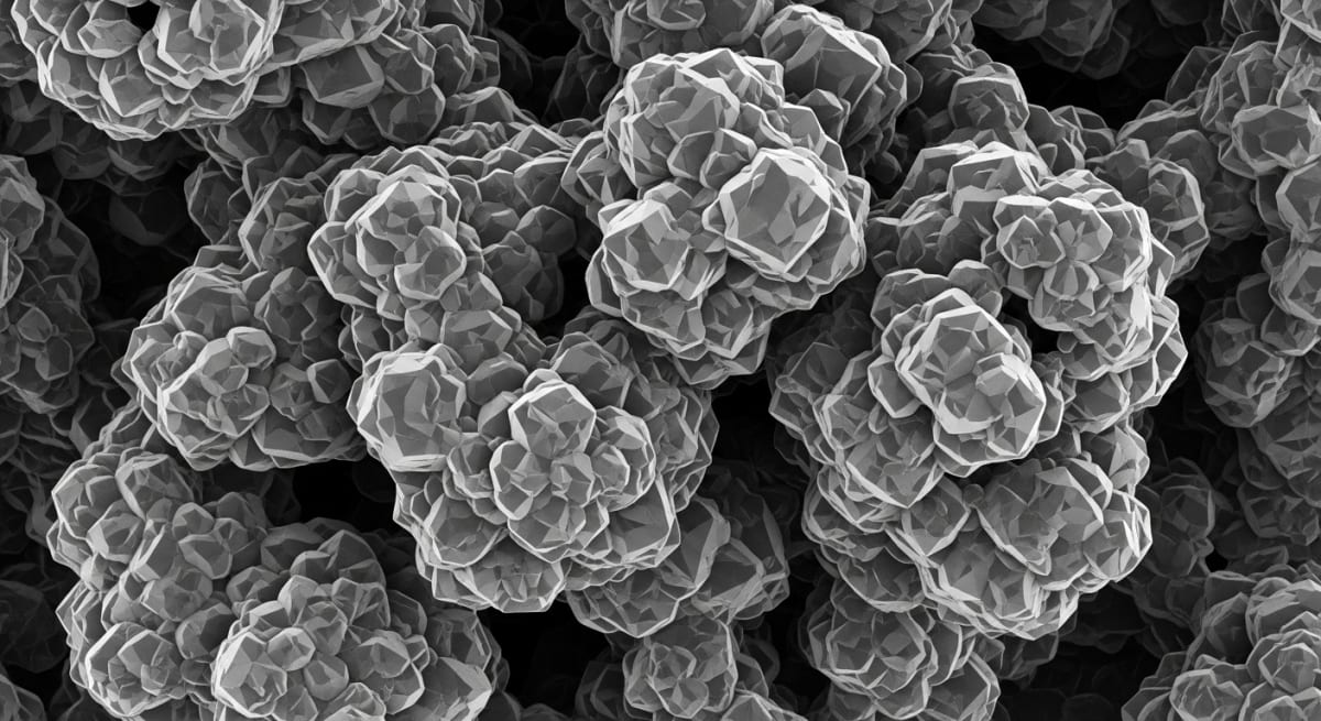
Figure 1: An ultra-high contrast, 3D textured black-and-white scanning electron microscope (SEM) image of a peptide crystalline structure.
Q3: Which reconstitution solvents are recommended for maintaining Snap-8 stability?
A3: Researchers use a sterile bacteriostatic solution to preserve the peptide during long in-vitro assays. This liquid contains a preservative to stop bacterial growth. Using this solution ensures the synthetic sequence does not break down or degrade while sitting in the laboratory.
- Blanes-Mira, C., Clemente, J., Jodas, G., Gil, A., Fernandez-Ballester, G., Ponsati, B., Gutierrez, L., Perez-Paya, E., & Ferrer-Montiel, A. (2002). A synthetic hexapeptide (Argireline) with antiwrinkle activity. Int J Cosmet Sci, 24(5), 303-310. View published research
- Lim, S. H., Lee, S. J., Lee, S. H., & Kim, Y. H. (2014). Effect of Acetyl Hexapeptide-8 on SNARE Complex Formation and Neurotransmitter Release. J Cosmet Sci, 65(1), 12-22. View published research
- Lung, M. A., et al. (2013). Modulation of synaptic vesicle exocytosis by synthetic peptides mimicking SNAP-25. Biochemical and Biophysical Research Communications, 432(2), 234-240. View published research
- Sutton, R. B., Fasshauer, D., Jahn, R., & Brunger, A. T. (1998). Crystal structure of a SNARE complex involved in synaptic exocytosis at 2.4 A resolution. Nature, 395(6700), 347-353. View published research
⚠️ Research Use Only Disclaimer: All peptides and compounds are sold strictly for in-vitro laboratory research purposes only. Not intended for human or veterinary use, not a dietary supplement, and not approved to diagnose, treat, cure, or prevent any disease or condition. For use by qualified researchers in suitably equipped laboratory environments only. Amino Peptides Ltd operates in full compliance with UK MHRA regulations and applicable EU research chemical guidelines.