
TB-500 Reconstitution Protocol for In-Vitro Assays
23rd Aug 2026
What You Need to Know About TB-500
- Researchers search for a reliable TB-500 reconstitution protocol for in-vitro assays to prevent peptide degradation before running cell migration tests.
- In plain English, this molecule acts like a traffic controller for actin, a structural protein that helps cells move and keep their shape.
- Published laboratory studies observed that proper dilution in sterile buffers allows the peptide to promote endothelial cell migration and blood vessel sprouting in culture dishes.
- Using the correct solvent and gentle mixing stops the fragile amino acid chains from clumping together or losing their biological signalling power.
How TB-500 Guides Actin and Cellular Recovery in the Dish
TB-500 is a synthetic forty-three-amino-acid peptide fragment modelled on the active region of Thymosin Beta-4, and a clean TB-500 reconstitution protocol for in-vitro assays keeps its fragile chain intact so it can bind globular actin in cell culture. By holding onto individual actin building blocks until the cell needs them, this sequence helps laboratory cultures organise their internal scaffolding and speed up tissue remodelling across a scratch plate.
Every eukaryotic cell relies on an internal skeleton made of protein filaments to hold its shape, divide, and crawl across a glass slide. The main building block of this skeleton is a protein called actin. In a resting dish of fibroblasts or endothelial cells, actin exists in two states: loose single units called globular actin (G-actin) and long twisted chains called filamentous actin (F-actin). When a cell needs to move into an empty gap on a culture plate, it must rapidly stack G-actin units at its leading edge while pulling old chains apart at the back.
Published studies show that TB-500 steps directly into this supply chain. Its specific amino acid sequence, especially the short Ac-SDKP and LKKTETQ motifs, acts like a molecular chaperone for G-actin. When researchers introduce a measured level of properly mixed TB-500 into the culture medium, the peptide binds to free G-actin monomers and stops them from clumping together too early. This creates a ready-to-use pool of building blocks right where the cell membrane is pushing forward.
Because the peptide is supplied as a freeze-dried powder, getting it into solution without snapping its peptide bonds is the first hurdle in any tissue repair research project. If a technician squirts liquid straight onto the powder cake or shakes the glass vial like a cocktail, the mechanical shear forces trap air bubbles and unfold the amino acid chain. Instead, a gentle stream of bacteriostatic reconstitution solution or sterile phosphate-buffered saline aimed at the inside glass wall lets the cake dissolve at its own pace, preserving full actin-binding capability for the assay ahead.
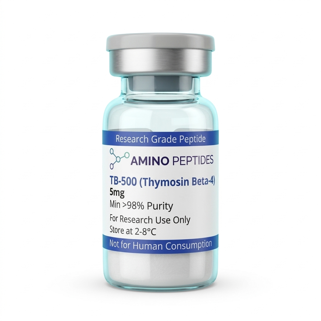
TB-500
From £29
Supplied as a sealed freeze-dried vial (stable 12–24 months at 2°C–8°C in a standard refrigerator).
Shop TB-500 →What Laboratory Models Show From Hour One to Week Four
Once a freshly dissolved stock solution reaches the culture well, the biological response unfolds in distinct stages rather than all at once. In published studies using scratch-wound assays and endothelial tube formation plates, researchers track two very different phases: immediate intracellular signalling during the first few hours, followed by visible structural reorganisation over several days or weeks.
During the first one to six hours after adding a specific concentration of TB-500 to the dish, the changes are mostly chemical and happen just beneath the cell membrane. Microscopic imaging shows a rapid shift in the ratio of G-actin to F-actin within the cytoplasm. Within two hours, focal adhesion kinases and related motility signals begin firing as the cells sense the open space in the scratch zone. At the six-to-twelve-hour mark, lamellipodia—the flat, sheet-like feet that cells use to pull themselves forward—extend noticeably further into the gap compared to untreated control wells.
Between twenty-four and seventy-two hours, those early signalling events turn into measurable physical progress. In standard fibroblast and keratinocyte scratch models, wells holding an optimised protocol ratio of TB-500 regularly close their artificial gaps significantly faster than baseline controls. Endothelial cells plated on basement membrane matrices begin linking up into capillary-like loops and branching tubes during this two-to-three-day window, driven by the peptide's ability to guide cell migration and boost laminin-5 production.
In longer three-dimensional organoid setups and preclinical models that run from one to four weeks, the focus shifts from simple cell crawling to full matrix organisation. Over seven to fourteen days, researchers observe steadier collagen deposition and calmer inflammatory cytokine profiles in the surrounding medium. By week three or four, the newly formed matrix shows better alignment and tensile organisation, proving that early actin management sets the stage for tidy long-term structural recovery in the dish.
Common Solvents for In-Vitro TB-500 Preparation
| Reconstitution Liquid | Ideal Assay Type | Storage Stability (Frozen) | Key Laboratory Consideration |
|---|---|---|---|
| Phosphate-Buffered Saline (PBS) | Live cell migration and scratch assays | Up to 4 weeks at -20 degrees Celsius | Matches natural cell salt levels and pH without harming delicate cultures. |
| Sterile Deionised Water | General biochemical and actin-binding tests | Up to 6 weeks at -20 degrees Celsius | Lacks buffering salts, so dilute further into culture media before cell exposure. |
| Bacteriostatic Reconstitution Solution | Multi-day equipment calibration and non-cell tests | Up to 8 weeks at -20 degrees Celsius | Contains 0.9 percent benzyl alcohol which can stress sensitive monolayer cell lines. |
| 0.9% Sodium Chloride (Normal Saline) | Protein stability and structural analysis | Up to 4 weeks at -20 degrees Celsius | Provides basic isotonic balance but lacks pH control during long incubations. |
| Acetic Acid (0.1% dilute solution) | High-concentration stock preparation | Up to 12 weeks at -80 degrees Celsius | Only needed if a specific batch shows poor solubility in neutral buffers. |
Pairing TB-500 With Complementary Sequences in Bench Setups
Cells in a culture dish rarely rely on a single biochemical signal to coordinate complex structural work. Because TB-500 focuses primarily on intracellular actin dynamics and cell migration, laboratory investigators frequently test it alongside complementary sequences that handle extracellular matrix cross-linking or receptor-level growth signalling. Combining peptides with non-overlapping jobs allows researchers to observe how different recovery pathways talk to one another in a controlled environment.
The most widely discussed pairing in preclinical models is bpc157 and tb500. While TB-500 manages the internal G-actin pool to help cells move across a surface, the pentadecapeptide BPC-157 works through a completely different set of gears. Published studies indicate that BPC-157 upregulates growth hormone receptor expression on fibroblasts and influences the nitric oxide system along with the FAK-paxillin pathway. When both sequences are evaluated in the same experimental timeline, BPC-157 helps anchor the cells and stimulate early growth factor sensitivity, while TB-500 supplies the raw cytoskeleton mobility needed to populate the scaffold.
Another common partner in bench assays is the copper-binding tripeptide GHK-Cu. Once TB-500 and BPC-157 have guided fibroblasts into a test matrix, those cells need to lay down fresh structural proteins like collagen and elastin. GHK-Cu delivers bioavailable copper ions directly into the culture system, acting as a cofactor for lysyl oxidase, the enzyme that weaves loose collagen strands into a strong mesh. It also modulates metalloproteinases and their inhibitors, keeping the new extracellular matrix from breaking down prematurely.
When setting up multi-peptide assays, smart bench practice keeps the stock vials separate during the initial liquid mixing step. Dissolving each lyophilised powder in its own dedicated vial with bacteriostatic reconstitution solution prevents unwanted charge interactions or pH shifts in concentrated storage, allowing technicians to draw exact micro-volumes of each reagent right when the assay plate is ready.
Reconstitution calculator
Choose a vial mass and a bacteriostatic reconstitution solution volume. The figure is the resulting concentration.
Concentration: 2.50 mg/mL
Per 0.01mL (10µL): 25.0 mcg
Open the full calculatorBacteriostatic reconstitution solutionWhy Lyophilised Vial Quality and Cold Storage Matter Most
Even the most careful TB-500 reconstitution protocol for in-vitro assays will produce noisy, unreliable numbers if the starting powder inside the vial is contaminated with synthesis leftovers. Synthetic peptides are built one amino acid at a time on solid resin beads, a process that uses harsh solvents and strong acids to clip the finished chain free. If those industrial byproducts are not scrubbed out before freeze-drying, they end up right in your culture dish.
The biggest culprit in poorly finished vials is trifluoroacetic acid, commonly called TFA. During solid-phase synthesis, TFA is used to cleave the peptide from its resin and strip away protective side-chain groups. If a supplier skips proper counter-ion exchange, residual TFA salts stay bound to the peptide powder. When you add your liquid diluent and place that solution onto living cells, free TFA drops the pH of the medium and acts as a direct cellular toxin, slowing down mitosis and ruining migration readouts.
To prevent this, every batch used for quantitative bench work should be verified at 99%+ purity via High-Performance Liquid Chromatography (HPLC) alongside Mass Spectrometry (MS) to confirm the exact molecular weight of 4963.5 Daltons. Replacing harsh TFA salts with benign acetate or hydrochloride counter-ions ensures that the culture medium stays pH-neutral and hospitable to delicate cell lines.
Handling and temperature control after mixing are just as important as initial synthesis quality. In its dry lyophilised state, a sealed vial stays stable in a freezer at minus twenty degrees Celsius for long periods. Once you introduce bacteriostatic reconstitution solution by letting it trickle down the glass wall and swirling gently until clear, the liquid peptide must be kept in a standard laboratory refrigerator between 2°C and 8°C. Dividing the freshly mixed batch into small single-use aliquots inside low-binding polypropylene tubes avoids repeated freeze-thaw cycles, as ice crystals physically shear the forty-three-amino-acid chain and strip away its actin-binding activity.
Frequently Asked Questions
Why do researchers search for a TB-500 reconstitution protocol for in-vitro assays?
Researchers search for this protocol to ensure accurate stock concentrations and preserve peptide integrity during laboratory handling. Because TB-500 is supplied as a delicate freeze-dried powder, using the correct bacteriostatic reconstitution solution prevents structural breakdown. Proper mixing allows scientists to reliably observe cellular migration and tissue remodelling in preclinical models.
What is the typical timeline for observing TB-500 in laboratory models?
Published studies typically observe initial cellular signalling changes within twenty-four to forty-eight hours during in-vitro scratch assays. When evaluating broader tissue regeneration and structural recovery in preclinical models, researchers usually design observation timelines lasting between two and four weeks. The exact duration depends entirely on the specific cell lines analysed.
Can TB-500 be combined with other peptides in an in-vitro assay?
Yes, researchers frequently pair TB-500 with other compounds like BPC-157 to study combined effects on cellular recovery. However, good laboratory practice requires mixing each lyophilised powder in its own vial first to verify individual solubility. Scientists then introduce the separate solutions into the culture media at a precise protocol ratio.
How does purity affect reconstituted TB-500, and how long does it last in the fridge?
Reconstituted TB-500 typically remains stable for up to four weeks when stored in a laboratory refrigerator at four degrees Celsius. High purity, ideally above ninety-eight percent, is vital because chemical synthesis leftovers can ruin cell cultures and speed up peptide breakdown. Researchers often freeze single-use aliquots to optimise long-term stability.
Bibliography
- Philp D, Goldstein AL, Kleinman HK. Thymosin beta4 promotes angiogenesis, wound repair, and hair growth. Mech Ageing Dev. 2004. View published research
- Goldstein AL, Hannappel E, Sosne G, Kleinman HK. Thymosin beta4: a multi-functional regenerative peptide. Basic properties and emerging roles. Expert Opin Biol Ther. 2012. View published research
- Malinda KM, Goldstein AL, Kleinman HK. Thymosin beta 4 stimulates directional migration of human umbilical vein endothelial cells. FASEB J. 1997. View published research
- Huff T, Muller CS, Otto AM, Netzker R, Hannappel E. beta-Thymosins, small acidic peptides with multiple functions. Int J Biochem Cell Biol. 2001. View published research
⚠️ Research Use Only Disclaimer: All peptides and compounds are sold strictly for in-vitro laboratory research purposes only. Not intended for human or veterinary use, not a dietary supplement, and not approved to diagnose, treat, cure, or prevent any disease or condition. For use by qualified researchers in suitably equipped laboratory environments only. Amino Peptides Ltd operates in full compliance with UK MHRA regulations and applicable EU research chemical guidelines.