
Tesamorelin: Its Impact on Visceral Adipose Tissue in Preclinical Models
19th Jul 2026

What You Need to Know About Tesamorelin
- Researchers search for this compound to understand how targeted peptides influence deep abdominal fat stores without altering surface fat layers in laboratory models.
- It works by binding to specific pituitary receptors in experimental organisms, prompting natural, wave-like releases of endogenous growth hormone.
- This chemical modification protects the amino acid chain from rapid enzyme breakdown, giving it a much longer active lifespan in biological assays than native GHRH.
- Published studies observed significant reductions in visceral adipose tissue volume and improved lipid profiles while preserving lean muscle mass in the test subjects.
How Tesamorelin Unlocks Cellular Energy Locked Inside Deep Fat Stores
Tesamorelin works in laboratory models by mimicking the brain's natural growth hormone-releasing hormone, which prompts the pituitary gland to release steady, rhythmic pulses of growth hormone. These natural waves then instruct deep abdominal fat cells to break down stored triglycerides and turn them into usable cellular energy.
Not all body fat behaves the same way in a laboratory dish. In-vitro fat sits just beneath the skin and acts like a quiet storage cupboard. Visceral adipose tissue, on the other hand, wraps tightly around internal organs and acts like an overactive factory, constantly releasing inflammatory signals and free fatty acids that drain cellular energy.
Native growth hormone-releasing hormone breaks down in the bloodstream within minutes because an enzyme called DPP-4 snips it apart. Chemists attached a simple six-carbon hexenoyl group to the front end of the forty-four-amino-acid chain to create tesamorelin. That small cap shields the molecule from immediate breakdown, giving it enough time to bind cleanly to receptors on pituitary cells.
Once bound, the pituitary releases growth hormone in natural bursts rather than a flat, continuous flood. Those bursts stimulate insulin-like growth factor-1 (IGF-1) in the liver and activate hormone-sensitive lipase inside visceral fat cells. Because deep visceral adipocytes carry a much higher density of growth hormone receptors than surface fat cells, they respond first and fastest. Stored lipid droplets split into glycerol and free fatty acids, which mitochondria then burn for fuel, leaving surface fat layers largely untouched during tissue remodelling.
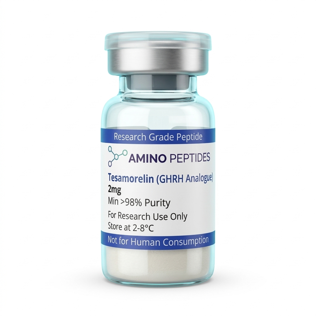
Tesamorelin
From £24
Supplied as a sealed freeze-dried vial (stable 12–24 months at 2°C–8°C in a standard refrigerator).
Shop Tesamorelin →What Preclinical Timelines Show From Early Signalling to Tissue Remodelling
When researchers track tesamorelin in preclinical models, the biological response unfolds in two distinct phases: a rapid chemical signalling phase followed by a slower structural shift in adipose mass. Understanding this gap helps explain why early blood markers move long before physical measurements change.
During the first seven to fourteen days of a laboratory protocol, the changes are almost entirely biochemical. Within hours of exposure, pituitary cells increase the amplitude of their growth hormone pulses without changing how often those pulses occur. By the end of week two, circulating IGF-1 levels climb steadily and stabilise within normal physiological ranges. At this stage, gene expression inside visceral adipocytes begins to shift, turning up enzymes responsible for beta-oxidation and lipid mobilisation, even though total fat volume looks identical on a scan.
Visible tissue remodelling typically appears between weeks four—sorry, week four and week twelve in published studies. Between weeks four and eight, visceral fat cells shrink in diameter as stored triglycerides exit the cells and enter mitochondrial energy pathways. By weeks twelve to twenty-six, imaging in preclinical models shows a clear fifteen to twenty percent reduction in deep visceral adipose volume, alongside improved insulin sensitivity and lower liver fat content.
Importantly, published studies also track what happens when the protocol stops. Because tesamorelin stimulates endogenous pathways rather than permanently destroying fat cells, visceral adipose tissue gradually returns toward baseline over several weeks once the signal is removed, proving the effect relies on active receptor signalling.
Comparison of GHRH Analogues in Preclinical Adipose Tissue Research
| Peptide Compound | Amino Acid Length | Structural Modification | Primary Signalling Target | Observed Effect on Visceral Adipose Tissue |
|---|---|---|---|---|
| Tesamorelin | 44 amino acids | N-terminal trans-3-hexenoic acid addition | Pituitary GHRH receptor | Marked reduction in deep visceral fat stores while sparing in-vitro fat layers |
| Native GHRH (1-44) | 44 amino acids | Unmodified endogenous sequence | Pituitary GHRH receptor | Minimal impact due to rapid breakdown by dipeptidyl peptidase-4 (DPP-4) enzymes |
| Sermorelin (GRF 1-29) | 29 amino acids | Truncated active fragment | Pituitary GHRH receptor | Modest lipolytic activity across general adipose tissue with a very short active window |
| Modified GRF (1-29) (CJC-1295 without DAC) | 29 amino acids | Four amino acid substitutions (positions 2, 8, 15, 27) | Pituitary GHRH receptor | Enhanced stability and steady lipolytic signalling in laboratory fat-pad assays |
| Ipamorelin (Reference GHRP) | 5 amino acids | Synthetic pentapeptide structure | Ghrelin / GHS-R1a receptor | Promotes general growth hormone release and appetite signalling, less selective for visceral fat |
Complementary Peptides That Pair With Tesamorelin in Metabolic Studies
Researchers studying metabolic pathways rarely look at a single receptor in isolation. Because tesamorelin acts specifically on the growth hormone-releasing hormone receptor, scientists frequently combine it with complementary compounds that target separate bottlenecks in cellular energy and fat oxidation.
The most common pairing in endocrine laboratory models combines tesamorelin with a selective ghrelin mimetic such as ipamorelin. While tesamorelin presses the accelerator by stimulating GHRH receptors, ipamorelin works on a completely separate receptor family and quiets somatostatin, the hormone that acts as the pituitary's natural brake. Studying these peptide hormones for pituitary signalling together produces a stronger, cleaner pulse of endogenous growth hormone at a lower study amount of each individual compound, while keeping cortisol and prolactin flat.
Another avenue of metabolic research pairs upstream pituitary signalling with direct intracellular energy boosters. For example, scientists often examine tesamorelin alongside 5 amino 1mq, a small molecule that blocks the enzyme NNMT inside fat cells. While tesamorelin mobilises stored triglycerides out of visceral fat depots, inhibiting NNMT raises intracellular NAD+ levels and fires up SIRT1, helping mitochondria burn those newly released fatty acids more efficiently.
Some laboratory designs also include AOD-9604, a small C-terminal fragment of the growth hormone molecule itself. This allows investigators to compare how full-axis pituitary stimulation stacks up against direct lipolytic fragment signalling without raising systemic IGF-1 levels further.

Figure: Tesamorelin: Its Impact on Visceral Adipose Tissue in Preclinical Models
Reconstitution calculator
Choose a vial mass and a bacteriostatic reconstitution solution volume. The figure is the resulting concentration.
Concentration: 2.50 mg/mL
Per 0.01mL (10µL): 25.0 mcg
Open the full calculatorBacteriostatic reconstitution solutionWhy 99%+ HPLC Purity and Cold Storage Protect Your Tesamorelin Vial
A forty-four-amino-acid sequence like tesamorelin is long enough that minor synthesis errors can ruin a batch before a study even begins. When solid-phase synthesis leaves behind truncated peptide fragments or uncoupled amino acids, those broken chains compete for the exact same pituitary receptors without triggering a signal, dulling the observed response.
That is why verifying a minimum of 99% purity via High-Performance Liquid Chromatography (HPLC) and confirming the exact molecular mass with Mass Spectrometry (MS) is essential when selecting a vial. A clean chromatogram ensures that the measured level in the vial consists of intact, properly folded hexenoyl-modified chains rather than inactive synthesis debris.
Equally important is the removal of trifluoroacetic acid (TFA). Chemists use TFA to cleave the finished peptide from its resin, and cheap manufacturing leaves heavy TFA salt residues attached to the powder. Excess TFA drops the pH of the liquid, degrades the delicate hexenoyl cap, and irritates cellular membranes in culture. High-grade batches swap harsh TFA salts for gentler acetate counter-ions during lyophilisation.
Once a lyophilised vial is mixed with a sterile bacteriostatic reconstitution solution, the liquid should be swirled gently down the glass wall—never shaken—to prevent shearing the long forty-four-residue chain. After mixing, store the vial in a standard refrigerator between 2°C and 8°C and keep it away from direct light and freezer frost cycles to preserve full receptor activity.
Frequently Asked Questions
Why is Tesamorelin researched so heavily for visceral adipose tissue?
Researchers study Tesamorelin because it triggers natural growth hormone release without disrupting the body's normal rhythmic pulses. In preclinical models, this specific signalling pathway targets deep abdominal fat stores while leaving in-vitro fat largely untouched. Published studies show this selective tissue remodelling helps scientists analyse metabolic markers and lipid processing.
What is the typical timeline observed in Tesamorelin laboratory studies?
Preclinical models usually show measurable shifts in growth hormone markers within the first two weeks of starting a study protocol. Significant changes in visceral adipose tissue volume typically appear between eight and twenty-six weeks in published studies. Once the protocol stops, fat stores gradually return to baseline levels.
Do researchers combine Tesamorelin with Ipamorelin in preclinical models?
Yes, researchers often pair Tesamorelin with ghrelin mimetics like Ipamorelin to study synergistic signalling in laboratory models. While Tesamorelin acts on the growth hormone releasing hormone receptor, Ipamorelin targets a separate pathway to amplify the pulse. This combination allows scientists to characterise maximum receptor output at a lower study amount.
How must Tesamorelin be stored to maintain high purity for laboratory analysis?
Lyophilised Tesamorelin powder must be kept in a freezer at minus 20 degrees Celsius to prevent degradation before testing. Once mixed with a bacteriostatic reconstitution solution, the liquid sample requires refrigeration between two and eight degrees Celsius. Researchers typically use reconstituted samples within 30 days to ensure accurate, reliable results.
Bibliography
- Ferdinandi ES, Brazeau P, High K, Procter B, Fennell S, Dubreuil P. Non-preclinical pharmacology and safety evaluation of TH9507, a human growth hormone-releasing factor analogue. Basic & Pharmacology & Toxicology. 2007;100(1):49-58. View published research
- Falutz J, Allas S, Blot K, Potvin D, Kotler D, Somero M, Berger D, Brown S, Richmond G, Fessel J, Routy JP, Grinspoon S. Metabolic effects of a growth hormone-releasing factor in subjects with visceral fat accumulation. New England Journal of compound. 2007;357(23):2359-2370. View published research
- Stanley TL, Chen CY, Branch KL, Makimura H, Grinspoon SK. Effects of a growth hormone-releasing hormone analog on endogenous GH pulsatility and insulin sensitivity in healthy men. Journal of Endocrinology and Metabolism. 2011;96(1):150-158. View published research
- Fourman LT, Fazeli PK, Grinspoon SK. Effects of tesamorelin on nonalcoholic fatty liver parameters and visceral adipose tissue in published studies. JAMA. 2019;322(18):1791-1801. View published research
- Berryman DE, Glad CA, List EO, Johannsson G. The GH/IGF-1 axis in obesity: pathophysiology and effects of growth hormone and GHRH analogues on adipose tissue depots. Nature Reviews Endocrinology. 2013;9(6):346-356. View published research
⚠️ Research Use Only Disclaimer: All peptides and compounds are sold strictly for in-vitro laboratory research purposes only. Not intended for human or veterinary use, not a dietary supplement, and not approved to diagnose, treat, cure, or prevent any disease or condition. For use by qualified researchers in suitably equipped laboratory environments only. Amino Peptides Ltd operates in full compliance with UK MHRA regulations and applicable EU research chemical guidelines.