
Glow vs Flash Bioluminescent Signal Emission in Cell Lysates
18th Nov 2026
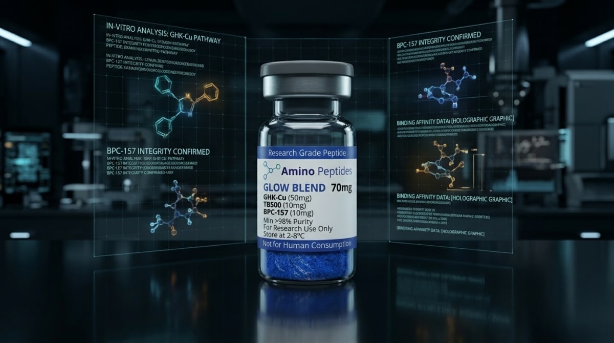
What You Need to Know About GLOW BLEND
- Researchers compare these two light emission styles to decide whether their laboratory plate reader needs automated liquid dispensers or can simply read stacked plates in large batches.
- Flash-type reactions create an immediate, intense burst of light that fades within seconds because the enzyme rapidly builds up inhibitory side products that block its own active site.
- Glow-type reactions add chemical stabilisers like coenzyme A and dithiol compounds to clear away those inhibitory byproducts, trading peak brightness for a steady light output lasting several hours.
- Published studies in mammalian cell lysates show that while flash assays offer raw sensitivity for single-tube measurements, engineered glow systems like NanoLuc deliver both high brightness and long stability for screening thousands of samples.
How Light-Emitting Assays Measure Cellular Energy and Peptide Activity
Bioluminescent signal emission in mammalian cell lysates acts as a direct optical speedometer for cellular energy and protein synthesis. By measuring light produced when luciferase enzymes react with their chemical substrates, researchers can see exactly how fast a peptide stimulates adenosine triphosphate (ATP) output or gene expression inside opened cells.
When curious readers look into the Comparative Characterisation of Sustained Glow-Type versus Flash-Type Bioluminescent Signal Emission in Mammalian Cell Lysates, they are usually trying to understand how laboratories actually prove a compound works. You cannot see cellular energy or collagen signalling with the naked eye. Instead, scientists break open the outer membranes of mammalian cells to create a rich soup called a lysate, and then use light-emitting reactions to count the active molecules inside.
The difference between flash-type and glow-type emission comes down to reaction speed and enzyme behaviour. Flash-type assays dump a raw substrate straight into the lysate, causing the luciferase enzyme to burn through its fuel all at once. This creates a blindingly bright burst of light that peaks within two seconds and fades away fast. It is ideal when a laboratory has an automated plate reader with built-in liquid dispensers that can measure a single well the exact instant the reaction starts.
Glow-type assays take a calmer, steadier approach. Chemists add special buffer ingredients, such as coenzyme A or slow-release chemical cages, that prevent the enzyme from choking on its own reaction by-products. Instead of a sudden spark, the lysate gives off a steady, gentle hum of light that lasts for thirty minutes to several hours. For scientists studying how peptides support cellular energy, mitochondrial ATP generation, or skin elasticity markers, glow-type kinetics allow them to test dozens of multi-well plates on a standard benchtop reader without worrying that the signal will die out before the machine finishes scanning the final row.
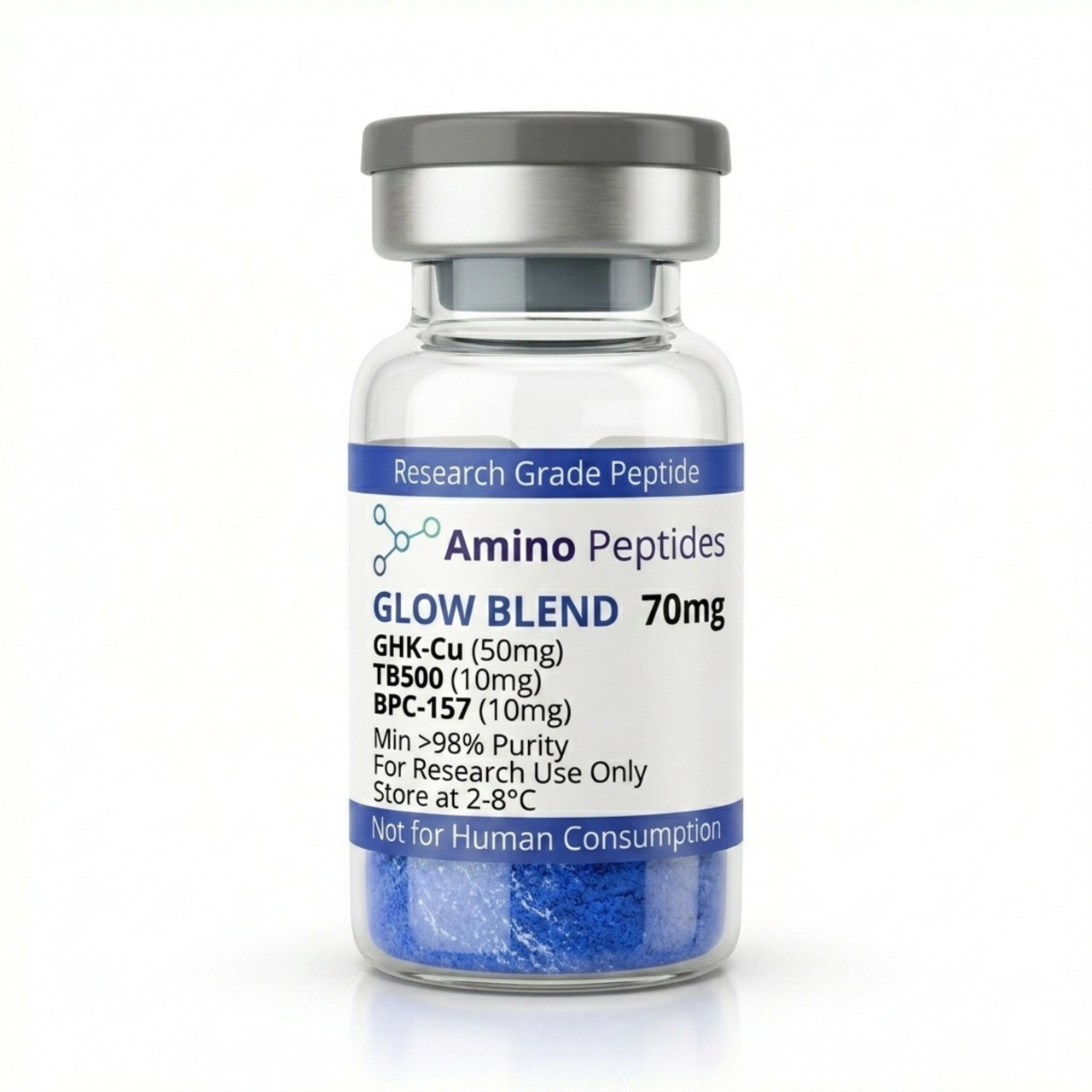
GLOW BLEND
From £65
Supplied as a sealed freeze-dried vial (stable 12–24 months at 2°C–8°C in a standard refrigerator).
Shop GLOW BLEND →Milliseconds to Hours: Tracking Early Signalling Versus Structural Remodelling
In laboratory models, choosing between a flash readout and a sustained glow readout depends heavily on the timeline of the biological event being measured. Peptide signalling does not happen all at once; it unfolds in distinct waves, starting with rapid electrical and chemical shifts and ending with slow, physical tissue remodelling.
During the first few seconds to minutes after a peptide contacts a cell culture, early signalling dominates. Receptors on the cell surface change shape, calcium ions rush across membranes, and secondary messengers like cyclic AMP spike sharply. When researchers lyse cells at these early time points, they often favour flash-type luminescence. Because early messenger molecules can be present in tiny, fleeting amounts, the massive photon burst of a flash reaction gives the highest possible sensitivity at a single snapshot in time. The light peaks between 0.5 and 2.0 seconds after mixing, decaying with a half-life of under ten seconds as oxyluciferin builds up and blocks the active site of the enzyme.
Later structural changes require a very different tracking strategy. When published studies investigate tissue repair research, they look at outcomes that take twenty-four to seventy-two hours to develop in culture dishes. These later milestones include fibroblast migration, fresh collagen type I and type III deposition, and sustained mitochondrial ATP expansion. To compare hundreds of samples collected across a three-day timeline, laboratories rely on glow-type emission.
By slowing down the turnover rate of the luciferase enzyme, glow reagents produce a flat, stable plateau of light with a half-life exceeding two to five hours. Although the peak brightness is lower than a flash burst, the steady output means a scientist can batch-process ninety-six or three hundred and eighty-four samples side by side. This stability ensures that a well read at minute five and a well read at minute forty-five report the exact same relative concentration of cellular ATP or reporter protein, giving a clean timeline of structural recovery.
Comparison of Flash-Type and Sustained Glow-Type Bioluminescence in Mammalian Cell Lysates
| Assay Parameter | Flash-Type Bioluminescence | Sustained Glow-Type Bioluminescence |
|---|---|---|
| Peak Signal Intensity | Very high initial burst of photons immediately upon mixing substrate with lysate | Moderately lower initial peak, optimised for steady photon output over time |
| Signal Half-Life | Short, typically decaying within 10 to 60 seconds due to rapid product inhibition | Extended, maintaining stable signal emission from 30 minutes up to several hours |
| Reagent Formulation | Basic buffer containing substrate, magnesium ions, and ATP without enzymatic turnover stabilisers | Formulated with coenzyme A, dithiothreitol, or proprietary enzyme inhibitors to prevent rapid self-inactivation |
| Hardware Requirements | Requires a luminometer equipped with onboard automated reagent dispensers for well-by-well reading | Compatible with standard batch-mode microplate readers and high-density automated stacking systems |
| Primary Laboratory Application | Low-throughput tube assays or ultrasensitive detection where maximum instantaneous photon count is critical | High-throughput screening of mammalian cell lysates across 96-well, 384-well, and 1536-well plates |
Pairing Multi-Peptide Stacks with High-Sensitivity Luminescence Readouts
Measuring one biochemical pathway in isolation rarely tells the whole story of how cells recover from stress. In preclinical models, researchers frequently combine complementary peptides to see if targeting multiple receptors at once produces a stronger, cleaner response in mammalian cell lysates. Bioluminescent assays are the primary way scientists verify that these combinations actually work together without exhausting the cell's energy reserves.
A popular example in laboratory investigations is the klow blend, which brings together four distinct sequences: GHK-Cu, BPC-157, TB-500, and KPV. Each component handles a separate job inside the culture dish. GHK-Cu delivers bioavailable copper ions that switch on genes linked to skin elasticity and extracellular matrix remodelling. BPC-157 accelerates growth factor receptor expression and nitric oxide balance, while TB-500 organises actin filaments so cells can physically move across a scratch in a culture plate. Meanwhile, the tripeptide KPV calms down inflammatory nuclear factor kappa B (NF-kB) signalling so the cells do not waste energy fighting false alarms.
When scientists test a four-part stack like this, they often set up multiplex luminescence assays inside the same lysate. For instance, they might use a glow-type firefly luciferase reaction to measure long-term collagen promoter activity driven by GHK-Cu and TB-500, followed immediately by a flash-type Renilla or NanoLuc reaction in the exact same tube to check rapid NF-kB suppression from KPV. Because the two enzymes use different chemical fuels and emit light at different wavelengths and speeds, the investigator gets two separate data points from a single sample. This dual-readout method proves how calming inflammatory noise frees up mitochondrial ATP, allowing the structural sequences to build new proteins much faster than any single peptide could manage alone.
Reconstitution calculator
Choose a vial mass and a bacteriostatic reconstitution solution volume. The figure is the resulting concentration.
Concentration: 2.50 mg/mL
Per 0.01mL (10µL): 25.0 mcg
Open the full calculatorBacteriostatic reconstitution solutionWhy 99%+ HPLC Purity and TFA Removal Protect Your Assay Readouts
Because bioluminescent assays can detect femtomole levels of activity, they are unforgiving when exposed to dirty reagents. If a flash or glow readout suddenly drops in brightness, the problem is rarely the mammalian cells themselves—it is almost always chemical contamination hiding inside a poorly manufactured peptide vial.
The biggest culprit in low-grade vials is residual trifluoroacetic acid, commonly known as TFA. During solid-phase synthesis, factories use TFA to cleave peptide chains off their resin beads. If the manufacturer skips proper salt-exchange steps, left-over TFA stays bound to the freeze-dried powder. When that powder is mixed with a bacteriostatic reconstitution solution and added to cell cultures, the harsh acid lowers the pH of the medium and damages cell membranes. Worse still, when the cells are lysed for luminescence testing, free TFA ions directly warp the three-dimensional fold of the luciferase enzyme. Even a tiny trace of acid can quench photon emission by thirty to percent, ruining both flash peaks and glow plateaus.
To avoid skewed data, experienced laboratories insist on batches verified at 99%+ purity via High-Performance Liquid Chromatography (HPLC) alongside mass spectrometry confirmation and dedicated TFA removal. Replacing toxic TFA salts with gentle acetate or hydrochloride counter-ions keeps the lysate pH right at its sweet spot of 7.4 to 7.8, allowing the luciferase reaction to shine at full strength.
Storage habits also directly protect assay accuracy. Lyophilised peptide vials and luciferin substrates should be kept in a dark refrigerator between 2°C and 8°C, or frozen at -20°C for long-term holding. Once reconstituted with a bacteriostatic reconstitution solution, keeping the vial chilled at 2°C to 8°C prevents thermal breakdown and oxidation, ensuring every measured level added to your plates delivers repeatable, interference-free light emission.
Frequently Asked Questions
Why do researchers search for comparative characterisations of glow-type versus flash-type bioluminescence?
Researchers compare these two assay formats to choose the best light-reading method for their mammalian cell lysate experiments. Flash assays produce a very bright burst of light for maximum sensitivity, while glow assays offer a steady, softer light that makes reading hundreds of laboratory samples much easier.
How does the study timeline differ between flash-type and sustained glow-type assays?
Flash-type assays require light measurement within seconds of adding the reagent, whereas sustained glow-type assays provide a stable reading window lasting from thirty minutes to several hours. This extended timeline allows scientists to analyse large batches of cell lysates without needing automated liquid dispensers inside their plate readers.
Can researchers stack or multiplex glow-type and flash-type bioluminescent reagents in one experiment?
Yes, scientists frequently combine different luciferase enzymes in the same mammalian cell sample to run dual-reporter assays. By pairing a flash-type reagent or a specific glow-type chemistry with an optical filter or quenching buffer, laboratory models can measure two separate cellular signalling pathways in a single test well.
How do reagent purity and fridge storage affect bioluminescent signal stability?
High substrate purity and strict cold storage are essential to prevent background noise and rapid signal decay in bioluminescent assays. Once mixed, most luciferase reagents lose activity if left in a standard four-degree fridge, so laboratories store aliquots at minus twenty degrees and avoid repeated freeze-thaw cycles.
Bibliography
- Hall MP, Unch J, Binkowski BF, et al. Engineered luciferase reporter from a deep sea shrimp utilizing a novel imidazopyrazinone substrate. ACS Chem Biol. 2012;7(11):1848-1857. View published research
- Thorne N, Inglese J, Auld DS. Illuminating insights into firefly luciferase and other bioluminescent reporters used in chemical biology. Chem Biol. 2010;17(6):646-657. View published research
- Fraga H. Firefly luminescence: a historical perspective and recent developments. Photochem Photobiol Sci. 2008;7(2):146-158. View published research
- Fan F, Wood KV. Bioluminescent assays for high-throughput screening. Assay compound Dev Technol. 2007;5(1):127-136. View published research
See our UK peptide research guides & COA briefings first in Google Search.
View published research⚠️ Research Use Only Disclaimer: All peptides and compounds are sold strictly for in-vitro laboratory research purposes only. Not intended for human or veterinary use, not a dietary supplement, and not approved to diagnose, treat, cure, or prevent any disease or condition. For use by qualified researchers in suitably equipped laboratory environments only. Amino Peptides Ltd operates in full compliance with UK MHRA regulations and applicable EU research chemical guidelines.