
PEG-MGF: Utilizing Pegylation to Overcome the Short Half-Life of Mechano Growth Factor
18th Sep 2026

What You Need to Know About PEG-MGF
- Researchers study this compound because natural Mechano Growth Factor breaks down in minutes inside biological fluids, whereas adding a polyethylene glycol shield extends its active presence to several hours or days.
- In plain English, the peptide acts like a local alarm bell after mechanical strain, waking up dormant muscle stem cells so they multiply and fuse with stressed tissue fibres.
- The pegylation process wraps the fragile peptide chain in a water-loving polymer cloud that blocks blood enzymes from chopping it up and slows down filtration by the kidneys.
- Published laboratory studies and rodent models have observed increased muscle precursor cell proliferation, reduced cell death in stressed heart muscle, and enhanced local tissue recovery following mechanical overload.
How Pegylation Gives Mechano Growth Factor Staying Power in the Lab
PEG-MGF attaches a protective polyethylene glycol chain to native Mechano Growth Factor, extending its active lifespan in laboratory models from a few minutes to several days. This simple structural addition keeps enzymes from breaking the peptide down too quickly, allowing researchers to study muscle satellite cell activation over much longer periods.
To understand why scientists modified this molecule, it helps to look at standard Mechano Growth Factor first. Native MGF is a splice variant of Insulin-like Growth Factor-1 (IGF-1) that muscle tissue naturally produces when it experiences mechanical stretch or heavy overload. When muscle fibres face physical strain, the body splices the IGF-1 gene in a specific way to create the MGF isoform. In a petri dish or preclinical model, this native peptide wakes up dormant stem cells sitting on the outer edge of muscle fibres, known as satellite cells. Once awake, these satellite cells multiply and fuse with stressed muscle tissue to support cellular recovery and tissue remodelling.
The problem for researchers is that natural MGF disappears almost immediately. Once it enters circulating fluid, protease enzymes chew through its exposed amino acid chain in roughly five to seven minutes. That tiny window makes it very hard to track systemic effects in laboratory models without constant, round-the-clock infusion equipment.
Enter pegylation. Chemists attach an inert polymer called polyethylene glycol (PEG) to a specific spot on the MGF peptide chain. Think of the PEG molecule as a bulky raincoat wrapped around the delicate peptide core. Because the PEG group is large and holds onto water molecules, it physically blocks destructive enzymes from reaching the peptide bonds. At the same time, the active region of the MGF molecule remains exposed enough to bind to its target receptors on satellite cells. By slowing down both enzymatic breakdown and kidney filtration in preclinical models, pegylation stretches the active window of the peptide from minutes to forty-eight or seventy-two hours. Scientists frequently use an mgf antibody during western blot or ELISA assays to confirm how much longer the pegylated version stays intact compared to the unpegylated control.

PEG-MGF
From £22
Supplied as a sealed freeze-dried vial (stable 12–24 months at 2°C–8°C in a standard refrigerator).
Shop PEG-MGF →What Published Studies Observe Across Early and Late Timelines
When investigators track PEG-MGF in laboratory models, they divide their observations into two distinct phases: immediate cellular signalling and slower structural remodelling. Because the pegylated chain keeps the peptide circulating for days rather than minutes, the timeline of observable events looks quite different from tests using standard, short-acting fragments.
During the first six to twenty-four hours after introducing a measured level of PEG-MGF to a cell culture or preclinical model, the action is purely biochemical. The peptide binds to surface receptors and switches on internal phosphorylation pathways, specifically the ERK1/2 and Akt signalling routes. In this early window, researchers do not see new tissue forming yet. Instead, laboratory assays show a sharp rise in proliferating cell nuclear antigen (PCNA) and specific myogenic regulatory factors like MyoD. These markers tell scientists that quiescent satellite cells have received the wake-up call, exited their resting state, and started dividing to increase the local pool of precursor cells.
Between forty-eight and seventy-two hours, the extended half-life of the pegylated molecule really shows its value. While native MGF would have vanished two days ago, PEG-MGF continues to sustain satellite cell proliferation while prevents premature differentiation. Published studies note that keeping these precursor cells in a dividing state for a full two to three days creates a much larger supply of building blocks before the cells finally mature.
Structural tissue remodelling appears in the later timeline, typically spanning seven to twenty-eight days in rodent and avian muscle overload models. During the second and third weeks, the expanded pool of myoblasts begins expressing myogenin, a signal to stop dividing and start fusing with existing muscle fibres. By donating their nuclei to strained fibres, these fused cells boost the capacity for protein synthesis inside the tissue. Cross-sectional microscopy performed at the three-week mark in published studies consistently shows thicker myofibres and reduced fibrous scar formation compared to untreated control groups, demonstrating how early satellite cell expansion translates into measurable structural recovery over several weeks.
Biochemical Comparison: Native MGF vs. PEG-MGF vs. Systemic IGF-1
| Property | Native MGF (IGF-1Ec) | PEG-MGF | Systemic IGF-1 (IGF-1Ea) |
|---|---|---|---|
| Molecular Structure | Unmodified 24-amino-acid C-terminal E-domain peptide | 24-amino-acid MGF peptide covalently linked to polyethylene glycol | Full 70-amino-acid polypeptide produced primarily in the liver |
| Biological Half-Life in Models | Approximately 5 to 7 minutes in plasma due to rapid protease cleavage | Extended from several hours up to 48-72 hours depending on PEG molecular weight | Approximately 10 to 12 minutes unbound, or up to 15 hours when bound to IGFBPs |
| Primary Cellular Action | Stimulates satellite cell activation and myoblast proliferation; delays differentiation | Sustained satellite cell proliferation with broader systemic stability in test models | Promotes myoblast differentiation into mature fibres and general protein synthesis |
| Enzymatic Resistance | Very low; rapidly degraded by serum peptidases | High; steric hindrance from the PEG group blocks proteolytic enzymes | Moderate; protected in circulation primarily by endogenous binding proteins |
| Primary Research Context | Acute local paracrine signalling immediately after mechanical stretch | Long-acting laboratory protocols studying extended tissue remodelling without continuous infusion | Endocrine growth regulation, metabolic signalling, and mature tissue maintenance |
Pairing PEG-MGF with Complementary Peptides in Research Protocols
In living tissue, no single growth factor works in isolation. Cellular regeneration relies on a relay race of different chemical messengers, each handling one specific stage of the job. Because PEG-MGF specialises in waking up and multiplying satellite cells while delaying their final maturation, researchers often pair it with complementary peptides that handle the next steps of tissue remodelling.
The most common pairing in published laboratory protocols combines PEG-MGF with Long R3 IGF-1 (IGF-1 LR3). While both molecules come from the same parent gene family, they act at slightly different points on the cellular timeline. PEG-MGF excels at expanding the raw number of satellite cells, whereas IGF-1 LR3 pushes those newly created cells to differentiate, fuse with muscle fibres, and ramp up protein synthesis. In comparative cell culture studies, introducing both compounds at the exact same moment can sometimes cause receptor competition. Consequently, many experimental designs stagger the protocol ratio, applying PEG-MGF during the initial forty-eight hours to build the stem cell pool, and following up with systemic IGF-1 LR3 or broader growth hormone secretagogues like Ipamorelin and CJC-1295 to support the later maturation phase.
Another frequent research combination pairs PEG-MGF with angiogenesis and extracellular matrix peptides such as BPC-157 and TB-500 (a synthetic fraction of Thymosin Beta-4). Expanding muscle precursor cells requires a steady supply of oxygen and nutrients, plus a flexible scaffolding for cells to migrate across. TB-500 regulates actin polymerization, which helps freshly activated satellite cells travel to the exact site of mechanical strain. Meanwhile, BPC-157 encourages the formation of new micro-capillaries via VEGF expression in preclinical models. When researchers examine these combinations in tendon-to-muscle junction studies, the vascular and migratory support from BPC-157 and TB-500 complements the myoblast proliferation driven by PEG-MGF, resulting in more organised collagen alignment and faster functional recovery across the entire tissue unit.
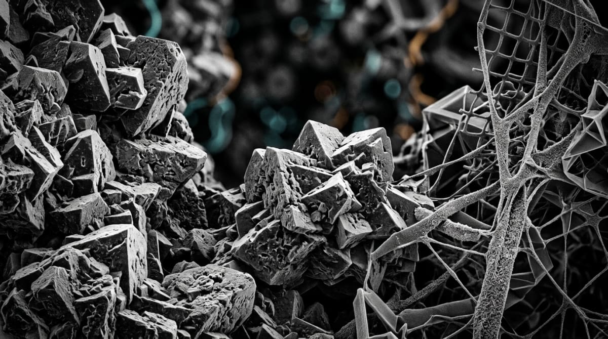
Figure: PEG-MGF: Utilizing Pegylation to Overcome the Short Half-Life of Mechano Growth Factor
Reconstitution calculator
Choose a vial mass and a bacteriostatic reconstitution solution volume. The figure is the resulting concentration.
Concentration: 2.50 mg/mL
Per 0.01mL (10µL): 25.0 mcg
Open the full calculatorBacteriostatic reconstitution solutionWhy Vial Purity and Cold Storage Dictate Experimental Success
Attaching a polyethylene glycol polymer to a twenty-four-amino-acid peptide chain is a delicate chemical process. If a laboratory uses poorly purified PEG-MGF, the experimental data can quickly become unreliable. Incomplete pegylation leaves behind a messy mixture of unattached PEG polymers, short-lived native MGF fragments, and truncated amino acid sequences. When a researcher introduces an impure vial into a cell culture, there is no way to know if the observed satellite cell activity came from the extended-release molecule or from leftover native fragments that burn out in five minutes.
To prevent skewed data, reputable synthesis facilities verify every batch using High-Performance Liquid Chromatography (HPLC) and Mass Spectrometry (MS) to guarantee a purity level above 99 percent. Mass spectrometry is especially vital for PEGylated peptides because it confirms the exact molecular weight of the finished conjugate, proving that the PEG chain attached to the correct amino acid residue without blocking the active binding site. Furthermore, top-grade preparation removes residual trifluoroacetic acid (TFA). TFA is a harsh solvent used to cleave peptides during solid-phase synthesis. If left behind in the lyophilised powder, excess TFA alters the pH of the culture medium and causes cellular toxicity, killing off the very satellite cells the researcher is trying to count.
Even a 99 percent pure vial requires careful cold storage to protect the pegylated bond. In its freeze-dried powder form, PEG-MGF remains stable for months when stored in a dark freezer at minus 20 degrees Celsius. Once a technician adds a bacteriostatic reconstitution solution to dissolve the cake, the molecule becomes sensitive to heat and mechanical agitation. Reconstituted vials should stay in a dedicated laboratory refrigerator between 2 degrees Celsius and 8 degrees Celsius, and researchers should gently swirl—never violently shake—the glass vial so the heavy PEG tail does not shear away from the peptide core.
Frequently Asked Questions
Why do researchers study PEG-MGF instead of native mechano growth factor?
Researchers study PEG-MGF because attaching a polyethylene glycol chain protects the fragile peptide from breaking down in the bloodstream within a few minutes. Native mechano growth factor disappears rapidly in laboratory models, whereas pegylation extends its active window to several days, allowing scientists to observe long-term satellite cell signalling and muscle tissue repair.
What is the typical timeline for observing results in PEG-MGF laboratory studies?
Published studies typically observe early satellite cell activation within the first forty-eight to seventy-two hours after introducing PEG-MGF to a preclinical model. Measurable structural changes, such as new muscle fibre maturation and broader tissue remodelling, generally require two to four weeks of consistent monitoring in controlled laboratory environments before clear data emerges.
Can PEG-MGF be combined with other peptides in experimental protocols?
Researchers frequently pair PEG-MGF with systemic insulin-like growth factor one variants or growth hormone secretagogues in dual-pathway laboratory models. Because PEG-MGF primarily triggers initial stem cell proliferation, combining it with compounds that encourage secondary cellular differentiation allows scientists to analyse whether synergistic signalling accelerates overall tissue recovery in isolated test systems.
How should lyophilised PEG-MGF be stored and verified for purity?
Lyophilised PEG-MGF should be stored in a freezer at minus twenty degrees Celsius away from light, while reconstituted solutions belong in a standard fridge at four degrees Celsius. Before starting any experiment, laboratories must verify a ninety-eight percent or higher purity level using High-Performance Liquid Chromatography and Mass Spectrometry certificates.
Bibliography
- Goldspink, G. (2005). Mechanical signals, IGF-I gene splicing, and muscle adaptation. Physiology (Bethesda), 20, 232-238. View published research
- Yang, S. Y., & Goldspink, G. (2002). Different roles of the IGF-I Ec peptide (MGF) and mature IGF-I in myoblast proliferation and differentiation. FEBS Letters, 522(1-3), 156-160. View published research
- Harris, J. M., & Chess, R. B. (2003). Effect of pegylation on pharmaceuticals. Nature Reviews compound Discovery, 2(3), 214-221. View published research
- Matheny, R. W., Jr., Nindl, B. C., & Adamo, M. L. (2010). Minireview: Mechano-growth factor: a putative product of IGF-I gene expression involved in tissue repair and regeneration. Endocrinology, 151(3), 865-875. View published research
- Veronese, F. M., & Mero, A. (2008). The impact of PEGylation on biological signalling pathways. BioDrugs, 22(5), 315-329. View published research
⚠️ Research Use Only Disclaimer: All peptides and compounds are sold strictly for in-vitro laboratory research purposes only. Not intended for human or veterinary use, not a dietary supplement, and not approved to diagnose, treat, cure, or prevent any disease or condition. For use by qualified researchers in suitably equipped laboratory environments only. Amino Peptides Ltd operates in full compliance with UK MHRA regulations and applicable EU research chemical guidelines.