
TB-500 (Thymosin Beta-4): An Analytical Review of Molecular Mechanisms and In-Vitro Applications
2nd Jul 2026

What You Need to Know About TB-500
- Researchers frequently investigate TB-500 and Thymosin Beta-4 to understand how isolated peptide sequences coordinate cellular movement and structural tissue remodelling in controlled laboratory environments.
- The peptide operates primarily by binding to globular actin (G-actin), which pauses filament clustering until cells need to build structural scaffolding and migrate across an in-vitro matrix.
- Published studies in preclinical models have observed accelerated blood vessel sprouting, elevated collagen deposition, and downregulated inflammatory cytokines during tissue recovery experiments.
- While Thymosin Beta-4 represents the complete 43-amino-acid naturally occurring protein, TB-500 typically refers to the synthetic research peptide or its central actin-binding fragment.
How TB-500 Guides Cellular Movement and Tissue Remodelling
TB-500 is a synthetic fraction of the naturally occurring Thymosin Beta-4 protein that laboratory studies use to examine rapid cellular migration and tissue remodelling. In isolated cell cultures, it works primarily by binding to globular actin, which helps cells build the internal scaffolding needed to move, divide, and organise new blood vessel networks.
Every living cell relies on an internal skeleton made of protein filaments to keep its shape and move across surfaces. The main building block of this skeleton is a protein called actin. Under normal conditions, cells keep a reserve pool of single actin units—known as G-actin—ready to snap together into long chains called F-actin. When a gap appears in a cultured tissue layer, nearby cells must quickly extend their edges and crawl into the empty space to close the breach.
Published studies show that Thymosin Beta-4 acts as the main regulator of this G-actin reserve. By holding onto single actin units until the exact moment they are needed, the molecule prevents premature clumping and allows the cell to extend smooth, directional projections. TB-500 focuses specifically on the active seven-amino-acid region (LKKTETQ) responsible for this actin-binding behaviour. Because TB-500 has a much smaller molecular weight than the full 43-amino-acid Thymosin Beta-4 parent protein, it diffuses through dense extracellular matrices in vitro with much less resistance.
Beyond simple cell movement, preclinical models demonstrate that this actin regulation triggers secondary signalling events. When endothelial cells migrate more efficiently, they begin to form capillary-like tubes, a process researchers call angiogenesis. At the same time, laboratory observations note a drop in pro-inflammatory cytokines within the surrounding culture medium. By calming local distress signals while speeding up physical cell travel, TB-500 gives scientists a clear model for studying how dense connective tissues—such as tendons, ligaments, and cardiac muscle layers—organise their structural recovery without forming excessive, stiff scar tissue.
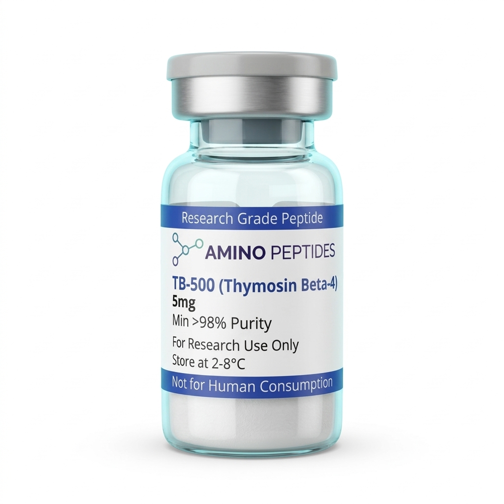
TB-500
From £29
Supplied as a sealed freeze-dried vial (stable 12–24 months at 2°C–8°C in a standard refrigerator).
Shop TB-500 →What Published Studies Observe Across Early and Late Phases
When researchers introduce TB-500 into an in-vitro assay or a preclinical model, the observed biological events unfold in distinct stages rather than all at once. Understanding the gap between immediate chemical signalling and slower structural changes helps explain how scientists design their observation schedules.
During the first 2 to 24 hours after exposure, the activity is purely biochemical and migratory. In scratch-wound assays—where a scientist scrapes a tiny line through a flat layer of cells in a dish—measured levels of Thymosin Beta-4 fragments cause border cells to polarise and begin creeping into the open gap within hours. Inside the cell, G-actin pools shift rapidly toward the leading edge. Simultaneously, early gene expression changes occur, downregulating acute inflammatory markers like nuclear factor kappa B (NF-kB) before any visible tissue architecture changes take place.
Between days 3 and 7 in extended culture models, the focus shifts from single-cell movement to group organisation. Endothelial cells that migrated during the first phase begin linking together to form early micro-vessels. Fibroblasts start laying down fresh, temporary extracellular matrix proteins, primarily type III collagen, to bridge larger structural gaps. In dermal and corneal laboratory models, this middle window is when surface closure rates visibly pull ahead of untreated control groups.
From week 2 through week 4 and beyond, published studies track macro-level tissue remodelling. Temporary matrix scaffolding gradually gets replaced by stronger, neatly aligned type I collagen fibres. Instead of the tangled, chaotic web of fibres typically seen in rapid scar formation, tissues exposed to Thymosin Beta-4 sequences in preclinical models show parallel fibre alignment that closely mimics original, uninjured tissue. Laminin-5 production also rises during this later window, anchoring newly formed surface layers firmly to the underlying basement membrane. Researchers monitoring long-term structural recovery therefore look past the quiet early days, knowing that physical tensile strength only appears at the end of the remodelling cycle.
Biochemical Comparison: Full-Length Thymosin Beta-4 vs. TB-500 (Active Fragment LKKTETQ)
| Molecular Attribute | Thymosin Beta-4 (Full-Length) | TB-500 (Synthetic / Active Fragment) |
|---|---|---|
| Amino Acid Sequence Length | 43 amino acids (complete endogenous chain) | 43 amino acids (synthetic full chain) or 7 amino acids (Ac-LKKTETQ fragment) |
| Approximate Molecular Weight | 4,963.5 Daltons | 4,963.5 Daltons (full synthetic) or ~889 Daltons (heptapeptide fragment) |
| Primary Biochemical Target | Monomeric globular actin (G-actin) sequestration and extracellular surface receptors | Direct binding to the G-actin subdomain via the conserved LKKTETQ motif |
| Observed In-Vitro Activity | Broad-spectrum cellular migration, endothelial sprouting, and anti-apoptotic signalling | Targeted actin polymerisation control and rapid diffusion across dense extracellular matrices |
| Laboratory Reconstitution | Soluble in sterile water, saline, or bacteriostatic reconstitution solution | Highly soluble in sterile water or bacteriostatic reconstitution solution at standard study amounts |
Pairing TB-500 With Complementary Peptides in Research Models
In living tissue models, cellular recovery rarely depends on a single pathway. Because TB-500 specialises in actin-driven migration and broad tissue mobility, researchers frequently pair it with complementary compounds that target entirely different bottlenecks in the regeneration process.
The most widely documented pairing in preclinical literature combines TB-500 with BPC-157, a fifteen-amino-acid gastric pentadecapeptide. While TB-500 manages the internal actin cytoskeleton to help cells travel across a culture dish, BPC-157 upregulates growth hormone receptor expression on fibroblasts and modulates nitric oxide pathways through the VEGFR2 receptor. In simple terms, TB-500 supplies the physical mobility to bring repair cells to the target site, while BPC-157 amplifies the local receptor sensitivity once those cells arrive. Tendon and ligament fibroblast models studying both peptides together consistently note faster matrix organisation than assays testing either sequence alone.
Another major research direction pairs TB-500 with copper-binding tripeptides for dermal and extracellular matrix investigations. Investigators who buy ghk-cu often examine how copper ions stimulate collagen and elastin synthesis alongside glycosaminoglycan production. In a combined study design, TB-500 accelerates the initial migration of keratinocytes and dermal fibroblasts across the test zone, while GHK-Cu drives the heavy structural synthesis of new collagen fibres and modulates metalloproteinases to clear out damaged proteins.
For studies looking at cellular stamina during heavy structural turnover, researchers also explore metabolic pairings. Building new actin filaments and secreting collagen requires enormous amounts of cellular energy in the form of ATP and NAD+. Adding a small-molecule NNMT inhibitor such as 5 amino 1mq allows investigators to observe how preserving intracellular NAD+ pools supports the high metabolic demand of migrating cells. By combining a structural mover like TB-500 with targeted receptor or metabolic enhancers, laboratory protocols can study multiple phases of tissue remodelling at the same time.
Figure: TB-500 (Thymosin Beta-4): An Analytical Review of Molecular Mechanisms and In-Vitro Applications
Reconstitution calculator
Choose a vial mass and a bacteriostatic reconstitution solution volume. The figure is the resulting concentration.
Concentration: 2.50 mg/mL
Per 0.01mL (10µL): 25.0 mcg
Open the full calculatorBacteriostatic reconstitution solutionWhy Vial Purity, TFA Removal, and Cold Storage Matter
Working with synthetic peptides in an analytical setting requires strict attention to how the raw material is manufactured, purified, and stored. Because TB-500 relies on a specific amino acid sequence to bind actin, even minor manufacturing leftovers in the vial can distort cell culture readouts.
When a lab syntheses TB-500, shorter broken chains—known as deletion sequences—can form alongside the target peptide. High-Performance Liquid Chromatography (HPLC) coupled with Mass Spectrometry (MS) is used to separate and verify the final product. A verified purity level of 99% or higher ensures that the measured level added to a petri dish consists almost entirely of active, intact peptide rather than inert fragments that compete for binding sites.
Equally important is the removal of trifluoroacetic acid (TFA). Standard solid-phase synthesis uses TFA to cleave the peptide from its resin, leaving behind TFA salts in low-grade batches. In sensitive in-vitro assays, residual TFA is toxic to living cells, alters the pH of the culture medium, and triggers false inflammatory signals that ruin migration data. Premium research vials undergo ion-exchange chromatography to swap harsh TFA ions for benign acetate salts, protecting delicate cell lines from acid stress.
Once a high-purity lyophilised powder arrives at the laboratory, temperature control keeps the sequence intact. Freeze-dried TB-500 vials should be stored in a dedicated refrigerator between 2°C and 8°C, shielded from direct light. When researchers prepare the powder for an assay using a sterile bacteriostatic reconstitution solution, the liquid must be introduced gently down the inside glass wall. Shaking a reconstituted vial creates foam and shear stress that can bend or snap peptide bonds. Gently swirling the vial until the powder dissolves, keeping the liquid refrigerated between 2°C and 8°C, and avoiding repeated freeze-thaw cycles ensures the molecular structure stays consistent from the first assay to the last.
Frequently Asked Questions
Why is TB-500 so widely searched in laboratory research?
Researchers search for TB-500 because it mimics the active region of Thymosin Beta-4, a protein involved in cellular tissue remodelling. Published studies show it binds to actin, a vital building block inside cells, which helps scientists analyse how cells move, divide, and support structural regeneration in laboratory models.
What is the typical timeline observed in TB-500 laboratory studies?
In preclinical models, researchers usually observe initial cellular signalling and migration shifts within 24 to 72 hours of introduction. Longer-term tissue remodelling and structural recovery markers in laboratory settings are typically analysed over a four-to-eight-week study window, depending on the specific concentration and protocol ratio used.
Why do researchers combine TB-500 with BPC-157 in in-vitro protocols?
Scientists often combine TB-500 and BPC-157 in laboratory models because they operate through different but complementary cellular pathways. While TB-500 directly regulates actin to support cell migration, BPC-157 influences growth factor signalling, allowing researchers to study whether combined protocol ratios optimise structural regeneration in cell cultures.
How should TB-500 be stored to maintain purity in the lab?
Unmixed, freeze-dried TB-500 powder remains stable for months when stored in a laboratory freezer at minus 20 degrees Celsius. Once mixed with a bacteriostatic reconstitution solution, the liquid compound must be kept in a standard fridge between 2 and 8 degrees Celsius and analysed within four weeks.
Bibliography
- Goldstein, A. L., Hannappel, E., Sosne, G., & Kleinman, H. K. (2012). Thymosin β4: a multi-functional regenerative peptide. Basic properties and preclinical applications. Expert Opinion on Biological signalling, 12(1), 37-51. View published research
- Philp, D., Goldstein, A. L., & Kleinman, H. K. (2004). Thymosin beta4 promotes angiogenesis, wound repair, and hair follicle development. Mechanisms of Ageing and Development, 125(2), 113-115. View published research
- Smart, N., Risebro, C. A., Melville, A. A., Moses, K., Schwartz, R. J., Chien, K. R., & Riley, P. R. (2007). Thymosin beta4 induces adult epicardial progenitor mobilization and neovascularization. Nature, 445(7124), 177-182. View published research
- Safer, D., Sosnick, T. R., & Elzinga, M. (1997). Thymosin beta 4 binds actin in an extended conformation and contacts both the barbed and pointed ends. Biochemistry, 36(19), 5806-5816. View published research
- Ho, E. N., Kwok, W. H., Lau, M. Y., Wong, A. S., Wan, T. S., Lam, K. K., Schiff, P. J., & Stewart, B. D. (2012). Doping control analysis of TB-500, a synthetic version of an active region of thymosin β4, in equine urine and plasma by liquid chromatography-mass spectrometry. Journal of Chromatography A, 1265, 57-69. View published research
⚠️ Research Use Only Disclaimer: All peptides and compounds are sold strictly for in-vitro laboratory research purposes only. Not intended for human or veterinary use, not a dietary supplement, and not approved to diagnose, treat, cure, or prevent any disease or condition. For use by qualified researchers in suitably equipped laboratory environments only. Amino Peptides Ltd operates in full compliance with UK MHRA regulations and applicable EU research chemical guidelines.